Featured Article
Please check out our Confocal Microscope section for more information or to find manufacturers that sell these products.
Late 16th century Dutch eyeglass maker Zaccharias Janssen and son Hans inadvertently invented the compound microscope. Since then, microscopy has enabled highly efficient and accurate molecular, genetic, and cellular imaging for countless research and clinical applications. Many forms of microscopes have been developed since Janssen and his son first discovered magnification through two convex lenses, but all can be divided into three categories: 1) optical, 2) electron, and 3) scanning tunnel microscope. Confocal microscopes are a type of fluorescence microscope and branch off from the optical microscopes category. This article will discuss the basic components of confocal microscopes, integral specifications, and key considerations necessary for selecting the best confocal microscope to suit the buyer’s needs.
Confocal microscopy background and features
Like all fluorescence microscopes, confocal microscopes use light to excite a fluorescent labeled specimen. Generally, the fluoresced light is a longer wavelength than the light source. Dichroic mirrors and emission filters matched to the spectral excitation and emission characteristics of the flourophore label are used to selectively image the fluorescence emitted by the specimen and not the light source. Figure 1 shows schematically how confocal microscopes use pinpoint illumination and sensing to dramatically reduce out-of-focus light as described by U.S. patent application 3013467, submitted in 1961 by Marvin Minsky.1
The advantages of confocal microscopes, as pointed out in the patent application, are as significant as they are numerous:
- Reduced blurring of the image caused by the scattering of light
- Increased resolution
- Improved signal-to-noise ratio (SNR)
- Permit clear examination of thick, light-scattering objects
- xy-scan and z-scan possible of wide areas of the specimen
- Electronic adjustment of magnification
- Especially well-suited for making quantitative studies of the optical properties of the specimen
- An infinite number of aperture planes in the microscope are potentially available for modulating the aperture with darkfield stops, annuli, phase plates, etc.
- Permit use of less complex objective lenses, including those for long working distance, ultraviolet, or infrared imaging, as they need to be corrected only for a single axial point.
The thin, clean, and clear optical sections made possible by using intense, monochromatic laser light sources make laser scanning confocal microscopy (LSCM) a natural selection for countless biological and medical science applications. Because LSCM allows for scanning living cells or biopsy samples at different focal depths, computerized reconstruction algorithms can assemble three-dimensional images of the specimen for nondestructive optical biopsy. Dynamic in vivo imaging is also possible. Composed of one or more electronic detectors, a computer, a multilaser system, wavelength selection devices, and a beam scanning platform, modern LSCM instruments are fully integrated electronic systems allowing for precise control of wavelength, excitation intensity, and depth of field.
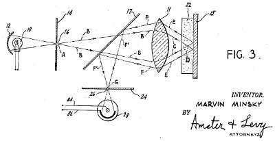 Figure 1 - Optical path of a confocal microscope. Excitation light pinhole, A, point D in the specimen, 22, and exit pinhole G are the focal points. A dichromatic mirror, 17, transmits beam A-B-C and reflects beam F’-G.
The primary advantage of LSCM is the ability to produce 0.5–1.5 μm serial optical sections through fluorescent labeled specimens. Using confocal microscopy principles, LSCM provides improved contrast and definition over widefield techniques, due primarily to a substantially better signal-to-noise ratio. Nondestructive sectioning enables both living and fixed-specimen examination without introducing artifacts commonly seen with physical sectioning and staining of specimens for traditional forms of optical microscopy.
Typically, spinning disk confocal microscopy (SDCM) uses two rotating disks with thousands of spatially arranged pinholes to selectively disperse excitation light across the sample. Rather than scanning the specimen one point at a time, SDCM is able to collect multiple fluorescent light points simultaneously while exposing the fluorophore-labeled sample to minimal excitation light. This may be critical when analyzing weakly expressing samples that are prone to photo-bleaching. The entire field-of-view is scanned in this manner during a single exposure, resulting in high-quality, high-speed confocal imaging.
Comparing single-point and spinning disk confocal microscopy
Image quality parameters such as overall efficiency of photon acquisition, image contrast, photo-bleaching susceptibility, and precision of quantitative measurement (based on pixel intensity) are useful in comparing confocal microscopy instruments, methods, or manufacturers (Table 1). Precision of image intensity is typically characterized as the SNR. In practice, the signal noise is generally attributable to the out-of-focus light saturating the detector and limiting the signal attainable from the in-focus plane.
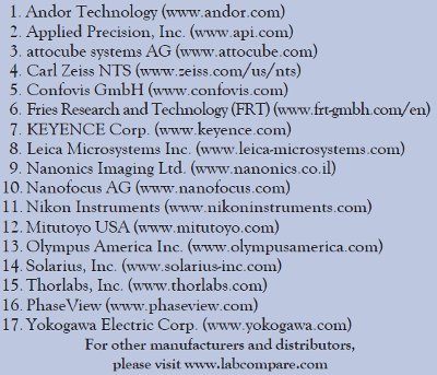
Table 1 - Confocal microscope manufacturers
Advantages of single-point over spinning disk confocal microscopes include a continuously adjustable pinhole diameter.2 When imaging specimens with out-of-focus fluorescence, the relatively large step or fixed pinhole diameter in SDCM will cause decreased SNR. Crosstalk between different pinholes on SDCM may begin to degrade contrast and SNR, depending on the thickness of the specimen and the spacing between pinholes.
However, SDCM collects approximately 103 pixels in parallel, as opposed to one pixel at a time collection, and exposes the sample to less excitation light, giving SDCM a large improvement in speed and higher resistance to photo-bleaching effects, respectively.
Ultimately, selecting between single-point and spinning disk confocal microscopy is dependent on application, specimen sensitivity to photo-bleaching, processing speed requirements, photon efficiency, contrast, and SNR needs.
Key purchasing considerations
To aid the buyer in understanding microscopy requirements, common confocal specifications and terms are defined below along with their respective implications.3
Measurement range: Confocal measurement range is limited by quantitative accuracy within a small contiguous subvolume of a specimen, coupled with finite source brightness, fluorescence saturation, and photo-damage effects to the specimen.
Numerical aperture (NA): The measure of the resolving power and light-gathering capacity of a lens given by the equation NA = nsinθ, where n is the refractive index and θ is the half-angle of light entering the lens. Essentially, as NA increases, the light-gathering capacity and resolution of the lens improve.
Depth of field: The range within which the specimen can be moved in the Z-direction while remaining in acceptable focus. Not to be confused with depth of focus; the distance in the Z-direction that information around a point in the sample is still in acceptable focus, which is dependent on the wavelength of the imaging source, aperture, and refractive index.
Optical resolution: The ability to distinguish two points or objects within a sample clearly. Generally quantified in micrometer X/Y separation or the distance between two points on the focal plane normal to the excitation light source.
Important price points of confocal microscopes
Confocal microscope buyers should not be swayed by the initial cost of the instrument. Each instrument should be evaluated in terms of what additional technical capabilities it may bring to the work flow in terms of cost per analysis and increased productivity. For instance, a more expensive microscope may have minimal maintenance or sample preparation costs, translating to a reduced cost per image. References from laboratories that own the current model can also be helpful when determining the reliability and ease of use of an instrument already integrated into the work flow of a laboratory.
Conclusion
Careful consideration of specific application requirements is essential to selecting the best confocal microscope to suit the buyer’s needs. Once understood and matched to a confocal microscopy method, additional considerations include the reputation of the vendor or manufacturer. Industry knowledge, depth of experience of the company, and other nonproduct issues should be evaluated in addition to technical needs.
References
- Marvin, M., Inventor. Dec. 19, 1961. Microscopy Apparatus. U.S. Patent US3013467.
- Murray, J.; Appleton, P. et al. Evaluating performance in the three-dimensional fluorescence microscopy. J. Microscopy2007, 228, 390–405.
- Inoue, S. Foundations of Confocal Scanned Image in Light Microscopy. In: Pawley, J. Handbook of Biological Confocal Microscopy, 4th ed.; Springer Science+Business Media, LLC: New York, NY, 2006; p 1.
T. Keith Brock, BS, is a Contributing Writer, American Laboratory/ Labcompare; e-mail: [email protected].
Please check out our Confocal Microscope section for more information or to find manufacturers that sell these products.