Featured Article

Please check out our Flow Cytometer sections for more information or to find manufacturers that sell these products
The ability to sort individual cells quickly based on their size or protein expression has had a significant, positive impact on any area of cell biology research or medicine in which a mixed population of cells needs to be sorted out. This is accomplished using flow cytometry, in which cells (or particles) in suspension are funneled single-file through a narrow opening that ends in a nozzle, such that droplets of fluid emerge one at a time. Each droplet may, or may not, contain one cell.
How does a flow cytometer function?
As the droplet falls, it passes through one or several lasers. If the cell is labeled with a fluorescent tag that is excited by the laser light, the fluorescent signal that it subsequently emits will be noted by detectors. The scatter of the laser light, as well as the fluorescent signal, tells a computer to which (prespecified) population each cell belongs. The computer directs the flow cytometer to send each droplet to the collection well for its group. For example, it may sort droplets into categories of no cell, cell with no fluorescent signal, cell with a green fluorescent signal, cell with a red fluorescent signal, and cell with both green and red fluorescent signals.
Researchers currently use flow cytometry for many complex applications, including:
- Immunophenotyping
- Apoptosis
- Cell proliferation
- Cell cycle regulation.
However, the range of powerful flow cytometry instruments available today could bewilder even the seasoned shopper of lab instrumentation. If you are looking for a flow cytometer, mulling over some key considerations will help you narrow your options (see Table 1).
Table 1 - Key considerations for choosing a flow cytometer

Considerations for purchasing a flow cytometer
Number of parameters to measure
The number of parameters you can measure simultaneously in one assay will depend largely on how many lasers and detectors your flow cytometer is equipped with. The wavelengths/colors of the lasers you use will dictate the excitation frequencies of the fluorophores that you can use. The possible number of fluorophore colors will also be determined by the number of detectors you use at the respective emission wavelengths. In addition, detectors for scattered light will provide estimates of the dimensions of the cells (or particles).
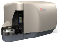
For example, the Gallios™ flow cytometer from Beckman Coulter (Indianapolis, IN; www.beckmancoulter.com) is equipped with up to 4 lasers and 10 detectors, allowing multicolor assays of up to 10 colors. Two lasers are the standard red and blue, with optional violet (405 nm excitation wavelength) and yellow (561 nm excitation wavelength) lasers also possible. The yellow laser can improve detection of red fluorescent proteins. Six fluorescence detectors are standard, with the option of adding 4 more, letting you assay 10 fluorescent signals simultaneously.
Throughput
The number of lasers you use will also affect how many ways you will need to sort the cells, for example, into 2, 4, or 6 sorted populations. The more sorted groups you have, the more you need to think about where to place them in your sample vessels. Typically, flow cytometers sort cells into sample tubes or microplates of up to 384 wells.
While it is possible to run single samples on most flow cytometers, the ability to use microplates makes higher throughput possible. Automation can augment throughput further still. Many variations and degrees of automation are available, from automated liquid handling systems that carry out particular sampling and dispensing jobs to fully automated robotic systems that execute all the functions of the work flow, including moving plates and samples around the flow cytometer. Deciding how much automation you want in your system can be tricky. On the one hand, reduced operator involvement removes human errors, and frees up additional human time for higher-level activities such as planning future experiments and data interpretation. On the other hand, because humans are more removed from the protocol, the experiment could potentially suffer from lack of valuable human judgment.
Sample volume
Other considerations include sample volume and cell counting. Most flow cytometers use sample sizes ranging from several microliters to hundreds of microliters. Also, some offer absolute cell counting features, while others reach cell counts using reference bead standards. For example, SPHERO™
Calibration Particles (Spherotech, Lake Forest, IL; www.spherotech.com) are reference beads that allow you to assess the sensitivity and linearity of the cytometer with your particular reagents.
Rare or precious samples
If you are using rare or precious samples, or trying to isolate a cell population of low abundance from a large mixture, you might want to look for particular flow cytometry solutions to these challenges. A preparatory purification or enrichment step prior to flow cytometry can make it easier to find your target cells. For example, Miltenyi Biotech (Bergisch Gladbach, Germany; www.miltenyibiotech.com) offers preenrichment options based on its MACS® technology, such as the MACSQuant® Cell Enrichment Unit, which can be integrated into the flow cytometry work flow for convenience.
Samples that are particularly vulnerable to environmental influences, or that require sterile conditions, might benefit from an entirely closed flow cytometry system, such as that offered by Partec (Münster, Germany; www.partec.com). Furthermore, adding another assay modality to your flow cytometer can help you derive the most information possible from each precious cell. If your experiments require fluorescence imaging during cell sorting, a flow cytometer with integrated imaging capabilities, such as the Amnis® Imaging Flow Cytometers’ ImageStreamX Mark II from EMD Millipore (Darmstadt, Germany; www.emdmillipore.com), may be useful. This instrument can take multiple images of each cell so that you can see the intracellular location of the fluorescence.
Another way to conserve precious resources is to use a much smaller volume with a microfluidics-based flow cytometry chip. This is like a flow cytometer in miniature. The microfabricated chip contains microfluidic channels into which a tiny sample is injected. Pneumatically controlled pumps and valves move the sample along the channels and single-file through laser(s) in a sorting area, where the scattered and fluorescent light is collected. Pumps accomplish the tailor-made movement instructions for individual cells—reroute the “red” cell to its group, the “green” cell to its group, and a “red and green” cell to its group. Microfluidic chips can boast lower background noise in general, but they have very low recovery rates (keeping in mind, of course, that the input sample was very small).
Alternatives to conventional systems
Creative instrument designers have gone beyond the conventional flow cytometer setup to offer interesting tweaks to the system. For example, the Attune® Acoustic Focusing Cytometer from Life Technologies (Carlsbad, CA; www.lifetechnologies.com) uses ultrasonic waves, rather than traditional hydrodynamic forces, to focus cells acoustically into a single-file line through a capillary. Its technology also lends greater accuracy and speed—it is up to 10 times faster than conventional flow cytometers, according to Life Technologies.
Another alternative system, the Guava EasyCyte™ flow cytometer from EMD Millipore, reads the sample from a microcapillary flow cell, rather than using hydrodynamic flow to move cells along through a sample area. Thus there are no droplets formed in these systems. Another example is the dropless CyFlow® Sorter from Partec, which incorporates a piezo valve moved by piezoelectric activation into the flow chamber. The piezo element directs each individual cell to its sorted destination.
Additional considerations
Additional factors might compete for your attention when looking at flow cytometers. These include:
- Size
- Interfaces
- Compatibility.
Today’s small benchtop models can be kept in an office, and portable models can be taken anywhere for field work. User-friendly interfaces and compatibility with existing automated machinery are also important features. Look for more flow cytometry innovations, too, as this important tool continues to advance quickly.
For more information on flow cytometers, please visit www. labcompare.com.
Also, see https://www.americanlaboratory.com/Blog/139016-The-Democratization-of-Cell-Sorting-Dont-Miss-the-Latest-Developments/
Caitlin Smith is a freelance science writer who has a Ph.D. in Neuroscience from Yale University and postdoctoral work in Electrophysiology and Synaptic Plasticity; e-mail: [email protected].
Please check out our Flow Cytometer sections for more information or to find manufacturers that sell these products