Please check out our Stereomicroscopes section to find manufacturers that sell these products
Stereomicroscopes are optical microscopes that give a three-dimensional (3-D) view of a sample and generally operate at low magnification. The 3-D image allows solid surfaces to be examined with greater clarity and permits the precise manipulation of samples during magnification.
A stereomicroscope effectively consists of two compound microscopes, each viewing the sample from a different angle. The stereoscopic image is achieved by maintaining two optical pathways through separate eyepieces focusing on the same point. This produces two separate images at a slightly differing angle to each other, one for each eye, enabling stereoscopic depth perception.
Stereomicroscopes are optical microscopes that give a three-dimensional (3-D) view of a sample and generally operate at low magnification. The 3-D image allows solid surfaces to be examined with greater clarity and permits the precise manipulation of samples during magnification.
Stereomicroscopes are used in food science, marine biology, embryology, environmental science, and forensic science. In pharmaceutical and biotechnology labs, stereomicroscopes are commonly used for magnifying biological samples such as plant tissues and organ samples. Wider industry applications include microsurgery, geology, and microelectronics, including the examination of circuit boards and microchips.
Considerations for purchasing a stereomicroscope
Stereomicroscopes, in common with all lab microscopes, have a wide range of functionality. At the most basic, a fixed magnification instrument with simple brightfield illumination is a good choice for the manipulation of large biological samples. At the other extreme, automated stereo zoom fluorescent microscopes with digital imaging allow for the time-elapsed recording of cell division and come with a wide range of accessories for other varied applications.
Stereomicroscopes are of the following main types:
- Common main objective (CMO) or Greenough design
- Fixed or zoom magnification.
Other specifications can be categorized by:
- Optics
- Illumination
- Ergonomics
- Stands
- Imaging
Types of stereomicroscopes
CMO or Greenough
 Figure 1 – The two main optical designs for stereomicroscopes: a) CMO design. b) Greenough design. (Image courtesy of Leica Science Lab by Leica Microsystems, Buffalo Grove, IL; www.leica-microsystems.com.)
Stereomicroscopes have two main optical designs: CMO, also known as the Galilean or telescope design, and Greenough (see Figure 1).
Common main objective microscopes have a single objective with two ocular channels and eyepieces. CMO microscopes can cost several times as much as Greenough microscopes, but they have superior light-gathering capabilities and higher resolution. Their modular design allows for the addition of a wide choice of accessories.
The Greenough design produces the stereo effect through the use of two angled objectives mounted side by side. Greenough microscopes are inexpensive and are ideal for everyday laboratory work.
Fixed or zoom magnification
Fixed magnification: The magnification is changed by swapping the eyepiece, either through replacement or by rotating a turret. Fixed magnification microscopes are the basic option. Lenses can be swapped to change magnification, for example, from 20× to 40×. When choosing a microscope, be realistic about the magnification level and range you require. For whole biological samples, the requirement may be as low as 10× or 20×. A typical working magnification range for a stereomicroscope is 10× to 100×.
Zoom magnification: The magnification can be altered on a continual basis. In zoom designs, the eyepieces can also be switched to change the range of magnification. For example, the M80 from Leica Microsystems has an 8:1 manual zoom, giving a magnification range of 7.5×–60×. Swapping lenses extends the magnification to 480×.
When choosing a zoom microscope, check the manufacturer’s zoom curves. To maintain optical quality, the lenses must remain correctly aligned during zooming. This is achieved with a precisely engineered mechanical zoom body. However, the SteREO Discovery.V20 from Carl Zeiss Microscopy (Jena, Germany) uses an electronic zoom body that calculates the optimal position of the lenses at each level of magnification.
The recently released SMZ25 from Nikon Instruments (Melville, NY; www.nikoninstruments.com) provides the world’s first zoom ratio of 25:1 (zoom range: 0.63×–15.75×), spanning spatial scales from single cells to whole organisms in one instrument.
Optics
Besides magnification, microscopes have many optical specifications that affect their suitability for different applications. These include:
- Working distance
- Depth of field
- Object field
- Numerical aperture and resolution
- Optical quality.
- Working distance: The distance from the bottom of the objective lens to the point of the sample in focus. Working distance is important since it affects the ease of manipulating the sample. However, working distance has an inverse relationship to numerical aperture (NA) and therefore resolution. At a large working distance of 171 mm, the SZX10® Research Stereomicroscope from Olympus (Center Valley, PA; http://www.olympus-ims.com/en/microscope) has an NA of 0.055; at 81 mm, the NA is 0.1, while at 33.5 mm it is 0.2.
- Depth of field: The distance between the nearest and furthest points from the objective that are in focus at the same time. When examining a very rough surface, a high depth of field is required. With flat samples, depth of field is not important. Depth of field is greater at lower magnifications.
- Object field: The diameter of the circular area of sample that is visible through the microscope at one time. Magnification and object field are inversely related, that is, at higher magnifications a smaller area of the sample is visible. Stereomicroscopes typically have large object fields to allow for sample manipulation.
- Numerical aperture: A unitless number derived by a complex formula that denotes the resolution of fine detail. For stereomicroscopes, the NA is dependent on the microscope objective and is not affected by eyepiece magnification. The higher the NA, the better the resolution, but, as described above, NA and working distance have an inverse relationship. Note that with all microscopes, the resolution varies with wavelength. Resolution is expressed as line pairs per millimeter (lp/mm).
- Optical quality: All optical systems have aberrations that vary with wavelength since light is refracted according to frequency. Optical instruments are designed to minimize these aberrations. Broadly, the methods of correction are:
a. Achromatic—useful when geometric shape is the priority
b. Apochromatic—useful when color is the priority
c. Plan—field curvature correction
d. PlanApo—apochromatic and flat field correction.
Illumination
Stereomicroscopes require illumination. Stereomicroscopes typically have two light sources: one from underneath the sample, and one above it. Incident light is commonly used for solid samples, and may be delivered using ring lights or spot lighting. Samples may be illuminated with UV or IR light sources in order to view specific characteristics or reactions. Illumination techniques include brightfield, darkfield, phase contrast, differential interference contrast, and confocal.
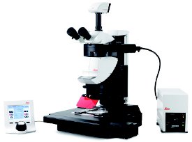 Figure 2 – The Leica M205 FA Automated Fluorescence Stereomicroscope combines apochromatic 20.5:1 zoom with Fusion Optics™ Technology that uses the right channel to produce very high resolution, while the left channel provides very high depth of field. (Image courtesy of Leica Microsystems.)
Working lifetime and versatility vary between different illumination systems. The BS-3060 Zoom Stereomicroscope from Carltex (Nyack, NY; www.carltex.com) has a light-emitting diode (LED) light for incident and transmitted illumination, with a life expectancy of up to 6000 hr.
Fluorescence stereomicroscopes, with magnifications in the region of 2500×, are widely used for cell biology, genetics, and embryology. The M205 FA Automated Fluorescence Stereomicroscope from Leica Microsystems (Figure 2) has a total magnification of 3.9× to 2560×.
Polarization contrast is widely used in plant biology. It reveals cellular structures, including cellulose and starch, without the need for staining.
Ergonomics
When choosing an instrument, consider whether it will be used by one person for long periods, or whether viewing angles, interpupillary distances, and working heights will need to be adapted to different users.
The Leica M80 has an adjustable 10° to 50° viewing angle for easy height adjustment. The interpupillary adjustment range for Motic Optical’s (Richmond, BC, Canada; www.motic.com) SMZ-171 microscopes is 48 mm–75 mm.
The Mantis Elite from Vision Engineering (New Milford, CT; www.visioneng.us) is designed so that the apparent distance to the viewed object image is identical to that of the real object, eliminating refocusing of the operator’s eye, thus reducing the likelihood of fatigue.
Stands
Your choice of stand will depend on the sample and the illumination technique. Check the flexibility and stability of different stands. The 1200ZB VanGuard Stereomicroscopes from VEE GEE Scientific (Kirkland, WA; www.veegee.com) (distributed by MIDSCI [St. Louis, MO; www.midsci.com]) feature a durable boom stand that permits 415 mm of horizontal travel and 300 mm of vertical travel and a solid base that requires no counterbalance.
Imaging
Trinocular heads add a digital camera to the two eyepieces, allowing for the recording of still or video images. The Olympus SZX16 has a tilting trinocular head that enables the user to switch between viewing and documentation.
Some digital imaging software will also record the optical settings for each image, making it easy to recreate the 3-D image as required.
Stereomicroscope manufacturers
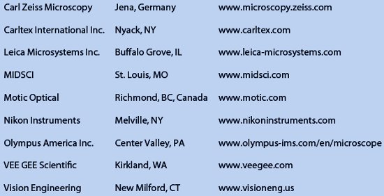
A list of stereomicroscope manufacturers is given in Table 1.
Table 1 – Manufacturers of stereomicroscopes
Katriona Scoffin, B.Sc., is a freelance science writer; e-mail: [email protected].
Please check out our Stereomicroscopes section to find manufacturers that sell these products