Featured Article

Following the successful labeling of antibodies with fluorescein isothiocyanate (FITC) by Albert Coons in 1941, new methods, instrumentation and an expanding menu of primary antibodies to targets of interest have led to increased applications of immunofluorescence (IF). Its utility, sensitivity and relative simplicity have made IF an essential tool for interpreting subcellular processes in a variety of fields such as drug-target identification and epigenetics. Options available for revealing cellular events using IF range from out-of-box multiplexed assay solutions to sophisticated image analysis software. The basic IF technique of labeling proteins with fluorescently conjugated antibodies, however, is characterized by a manual, repetitive liquid-handling workflow. This work automated a typical IF workflow to reduce manual labor, time and reagent consumption during an evaluation of oil-immersion microscope objectives.
Oil immersion is a common method in IF microscopy because it enhances optical resolution. This is of particular value when investigating small intracellular targets. IF was used to identify the subcellular location of organelle-specific proteins as a validation model for the system described in Ref. 1. Localization of these proteins can provide valuable information about their role in a variety of cellular processes, for example, determining the subcellular position of a marker that co-localizes with an organelle-specific antibody, or identifying a disease based on where distinct proteins occur. Expression of these biomolecules can vary in different cells and tissues, including their presence, size, and/or abundance. Assay optimization of cell types, antibody dilutions, direct and indirect labeling sensitivity and reagents are therefore important to successful results.
 Figure 1 – The MultiFlo FX Multi-Mode Dispenser (BioTek Instruments, Inc., Winooski, Vt.) can replace up to five reagent dispensers in a small footprint. The instrument was used in a cell- culture hood to automate sample preparation and immunostaining of an IF subcellular localization assay to validate oil-immersion objectives.
The liquid-handling steps of the sample preparation and immunostaining protocols available with the Cellular Localization IF Antibody Sampler Kit (Cell Signaling Technology, Danvers, Mass.) were automated (Figure 1). This facilitated scaling up to higher throughput beyond single microscopy slides, allowing the assay of multiple IF assay optimization parameters in parallel. The oil-immersion imaging technique validated here imposed two constraints that are important to consider when designing an automated liquid-handling profile as they directly influence vessel choice. First is the refractive index of the imaging vessel, which should match that of the oil column between the objective and the specimen. This serves to maximize light collection allowed by the objective’s numerical aperture, while preserving imaging resolution. Second, the bottom thickness and x,y,z physical dimensions of an imaging vessel can affect sample resolution and impede available imaging area due to the short working distance typical with oil objectives. Tolerances for resolution are included with immersion oils and objectives to aid vessel choice, and, for most automated imagers, vessel dimensions defined by the manufacturer are configurable in the software, allowing assessment of potential collision areas in advance.
Vessels that are optimal to the oil objectives tested are those with imaging surfaces that are 0.17 mm thick with a refractive index of 1.5 (the same as a #1.5-glass coverslip), with an 80–100% viewable area when using a standard ANSI/SLAS footprint vessel. Compatible vessels come in many shapes and sizes to allow a variety of throughput options. Vessel choice may be limited, however, by the area available for applying coverslips (and is dependent on the application), and the accessories available on a given liquid handler. Instruments that include stage adapters, multiple dispensing ports or dispense and aspirate manifolds of different sizes are useful. Dedicated reusable and autoclavable reagent-dispensing options reduce waste and cross-reactivity, while also making routine protocols easier to set up and maintain.
If standard instrument accessories limit protocol adaptation, users should consider automating subroutines, such as replacing the manual multiple wash steps common in immunostaining. Software features that allow customization of aspirate and dispense heights, flow rates, reagent volumes and offset values can enhance instrument performance. For example, when working with adhered cells, angled manifold tips with custom programmed offset values and flow rates can minimize cell loss from repeated rinse steps. Once an automated method is defined and validated, the scheduling routine can result in reagent savings and experimental reproducibility. Two or more vessels run successively, for instance, require only the same reagent overage volume as a single vessel. The right combination of accessories, instrument features and scheduling can provide optimal automation of an entire IF workflow, from sample preparation through mounting.
To demonstrate this, an automated liquid-handling IF protocol developed for a high-throughput microplate washer/dispenser in 96- and 384-well microplates2 was adapted to lower throughputs more conducive to oil-immersion imaging of coverslipped cells using a flexible multimode reagent dispenser. In one experiment, paraformaldehyde and methanol fixation methods were compared using eight antibodies in two multiwell vessels to determine if the antibodies were compatible with both methods. Methanol fixation requires less reagent preparation than paraformaldehyde and can be stored ready-to-use at –20 ºC.
The multimode dispenser was equipped with two syringe pump dispensers (fixatives), a 24-well interchangeable wash manifold (phosphate-buffered saline) and two peristaltic dpump dispensers, and was then placed in a cell-culture hood. Four 5-μL eight-tube autoclavable peristaltic pump cassettes were used. One was adapted with truncated dispense lines in single-tube mode, allowing eight unconjugated antibodies to be run in parallel using low active volumes; another was dedicated to cell seeding; and the other two were shared between blocking buffer (day 2) and two fluorochrome-conjugated secondary antibodies (day 3). The purge feature on the pumps permitted complete recovery of all unused antibodies and blocking buffer, in addition to enabling the eight primary antibodies to be dispensed from a single cassette with no cross-contamination. Automated assay was performed in two 24-well glass-bottom microplates (cat. no. P24G-1.5-13-F, MatTek Corp., Ashland, Mass.) using low flow rates and custom offsets to the lower left corner of each well. At the end of the protocol, mountant (Prolong Diamond Antifade Mountant [Invitrogen, Carlsbad, Calif.] with DAPI, cat. no. P36962 [Thermo Fisher Scientific, Waltham, Mass.]) and coverslips were applied manually to the bottom of the wells. Some manual intervention was also required at the primary antibody dispense step to switch tubes on the cassette from one set of four antibodies to another (see Figures 2–4).
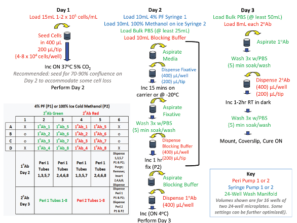 Figure 2 – Automated workflow of an IF protocol run during validation of oil-immersion objectives on an automated imager.
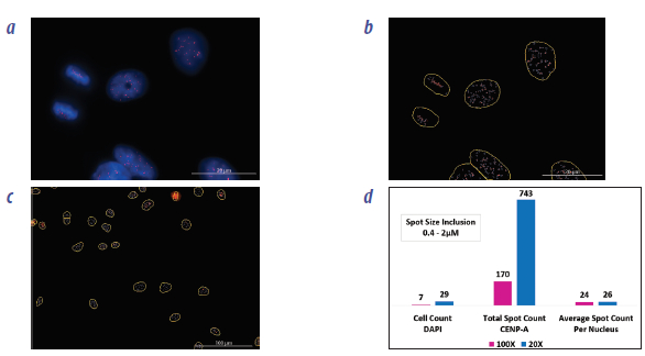 Figure 3 – Image analysis of HeLa cells following an automated liquid-handling workflow. a) CENP-A antibody (red) is targeted to centromeres, visible as small dots often <1 μM in size localized to the cell nucleus (DAPI counterstain, blue). b) A “spot counting” tool allowed scoring the expression of these subnuclear proteins (centromere spots, purple mask line; nucleus, yellow mask line). Due to their small size, high-resolution oil immersion (100×) was used to enhance spot threshold and size parameter optimization. c) Settings from the high-magnification analysis were applied to quantify spots in larger cell counts at low magnification (20×) and d) results were compared.
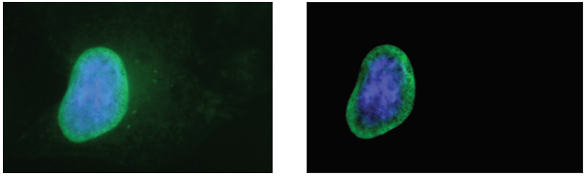 Figure 4 – Oil-immersion analysis (100×) of HeLa cells following an automated liquid-handling workflow. C39A3 antibody (green) detects endogenous levels of total NUP98 protein, a component of the nuclear envelope pore complex (DAPI nuclear counterstain, blue). Some non-specific binding and/or autofluorescence is common in IF. Software imaging tools like preprocessing can automatically adjust background and refine target thresholding as shown here before (left) and after (right) correction. Enhanced image quality and increased contrast of specific binding events can aid both qualitative confirmation and quantitative analysis of subcellular protein localization.
Conclusion
Automated liquid-handling instruments are well-suited to the preparation and labeling of cells for immunofluorescence. Vessels designed with a refractive index compatible with oil immersion make the technique amenable to automated immunostaining. Software features that aid in quantitative cellular analysis can add value to qualitative high-resolution confirmation of events. Affordable liquid handlers and combination imagers are available to complement most laboratories utilizing IF.
References
- www.biotek.com/resources/articles/oil-objective.html
- www.biotek.com/resources/articles/automated-tissue-culture-cell-fixation-staining.html
Wendy Goodrich is an applications scientist, BioTek Instruments, Inc., 100 Tigan St., Winooski, Vt. 05404, U.S.A.; tel.: 888-451-5171; e-mail: [email protected]; www.biotek.com. All image analysis was done on a Lionheart FX with Gen5 3.03 Image Prime software (BioTek Instruments, Inc.). Primary antibodies and fluorochome conjugated secondary antibodies (Alexa Fluor 594 and Alexa Fluor 488) courtesy of Cell Signaling Technology (Danvers, Mass.). AlexaFluor is a registered trademark of Life Technologies Corp. (Thermo Fisher Scientific, Waltham, Mass.) and dye antibody conjugates are sold under license from Molecular Probes (Eugene, Ore.).