Nanomaterials take biosensors to really small sizes
Not everyone agrees on what makes something a nanomaterial, except that some aspect of it is really small—nanometer scale. That doesn’t mean something must be just a millionth of a meter to make a material “nano,” but some aspect of it must be less than 100 nanometers in some dimension, according to the definition used by the U.S. National Institute of Environmental Health Sciences. Whatever the standard for a nanomaterial, it can’t be seen, not even with an ordinary lab microscope. It’s their smallness that makes nanomaterials particularly useful in building biosensors.
At the University of Connecticut (Storrs), Yu Lei, professor of chemical and biomolecular engineering, used titanium dioxide (TiO2) nanofibers to make a biosensor that detects nerve agents—such as paraoxon, methyl parathion and parathion—in organophosphate pesticides. Lei says that he and his colleagues made the biosensor from “a functional nanocomposite which consists of elastin-like-polypeptide-organophosphate hydrolase, bovine serum albumin, titanium–dioxide nanofibers and carboxylic acid functionalized multiwalled carbon nanotubes.” The TiO2 nanofibers, Lei explains, “enrich organophosphates in the nanocomposite due to its strong affinity with the phosphoric group in organophosphates, while carbon nanotubes were used to enhance the electron transfer in the amperometric detection as well as for covalent immobilization of the hydrolase.” The result was ultra sensitivity.
The nanomaterial used depends on what will be measured. At the University of Florida (Gainesville), Charles Martin, professor of chemistry, makes biosensors with single pores, which can be as small as 2 nanometers across, in polymer films. “This nano-scale size is critical to the sensor design, because—in one embodiment of the concept—we simply count protein molecules as they move through the pore,” Martin says. “This is accomplished by passing an ionic current through the pore and measuring the transient blockage of that current as the protein moves through the pore.” To measure a blockage in the current, the pore and whatever is going through it—a protein in this example—must be about the same size.
To measure small things, like biological components, miniaturized sensors can deliver bigger and more specific signals, and that makes nanomaterials perfect platforms for biosensors.
Building a biopsy
Nanomaterials really benefit clinical applications of biosensors, such as applying them to cancer diagnostics. Even an effective cancer treatment can fail at some point, and this often is the result of mutations. Various cancers depend on a mutation in the KRAS gene. In one of these mutations, an aspartate amino acid replaces a glycine at position 12 in KRAS—a so-called KRAS G12D mutation. Andrew Ko, professor of medicine at the University of California, San Francisco, is using technology from Two Pore Guys (Santa Cruz, Calif.) to see if he can detect KRAS G12D mutations in cell-free, circulating tumor DNA (ctDNA).
A simple method to pick up this mutation can be used in many ways. For a patient in cancer remission, this mutation could reveal some of the earliest signs of cancer recurrence. It could also be used to study the efficacy of a treatment. All of this could be done most efficiently with a liquid biopsy using bodily fluids such as blood or urine.
The technology from Two Pore Guys uses a 30-nm-thick piece of silicon nitride (Si3N4) with a 25–30-nm pore inside it. Placing this Si3N4 substrate between two chambers of fluid leaves only one path between them, and that’s the pore. “When voltage is applied, negatively charged molecules pass through the pore, impeding the current by a measured amount,” says Dan Heller, CEO at Two Pore Guys. “Individual molecules are thereby counted.” He adds, “This is old science that has been known since 1999.”
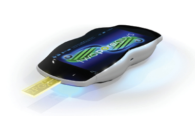 Using a nanosize pore and molecular methods, this device counts specific target molecules. (Image courtesy of Two Pore Guys.)
The novelty of the Two Pore Guys technology comes from adding substantial molecules to specific targets, such as proteins or DNA. “When those targets go through the pore, they produce uniquely larger measurements than all other molecules,” Heller explains. “This now provides the apparatus, whereby sample fluids of any type can be input, so that when they mix with the target-specific chemistries and the voltage is applied, all the targets can be counted, and the non-targets—background—are ignored.”
The result is a nano-based biosensor that detects single, specific molecules in a sample that requires little preparation. Heller adds that these biosensors “can be mass-produced at scale at very low cost, and can be incorporated into inexpensive, simple devices that anyone can use.”
 Carbon nanotubes (gray mesh) and various other forms of nanomaterials can interact with biological elements, such as H5N2 avian flu virus (purple rods) shown here, and that opens many approaches to building biosensors. (Image courtesy of Penn State University, State College.)
Tracking the technology
Although most microscopes cannot image nanomaterials, some instruments, such as PerkinElmer’s (Waltham, Mass.) IVIS in vivo imaging systems, can. A scientist can tag nanomaterials with fluorescent labels and use this platform to study where the nanomaterials go in an animal, such as a mouse or rat. This can be used to “study particle distribution, residence time in the host and targeting efficiency to specific organs,” explains Vivek Shinde Patil, senior manager, technical applications, preclinical imaging at PerkinElmer. “Noninvasive imaging also allows for studies of nanomaterial toxicity and to highlight their utility as biosensors to detect hypoxia, inflammation and a wide repertoire of pathologies.”
Imaging can help scientists decide which nanomaterials to use in a particular biosensor. One could try different nanomaterials and see where they go and how long they stay there. Optical properties of a nanomaterial might even matter. “For instance, researchers have used gold nanoclusters or nanorods for detection of plaques in Alzheimer’s disease because of their interesting optical properties,” Patil says.
For clinical biosensors in general, imaging can be used to improve a nanomaterials-based device. “The IVIS has been used preclinically to characterize nanomaterial biosensing utility in areas such as oncology, inflammation and neurodegeneration,” says Patil. “For example, an IVIS user has developed biocompatible hypoxia sensors using photonic nanomaterials.” In that application, the level of oxygen triggers a wavelength shift in the fluorescence of the particles, which distinguishes pre- and post-hypoxic conditions. As Patil points out, “Imaging offers unique and novel insights into nanomaterial behavior and function.”
These examples of nano-based biosensors reveal the variety of technologies that can be used, from microscopic pores to innovative imaging. The component being measured and where it is measured determines the best combination of a nanomaterial and a method of sensing. Its popularity will also be impacted by the ability to manufacture and use the biosensor. One thing’s certain: nanomaterials help biosensors get small, really small.
Mike May is a freelance writer and editor living in Florida. He can be reached at [email protected].