Featured Article

Fourier transform infrared (FTIR) microspectroscopy is a preferred method for the analysis and identification of contaminants, fibers, and other microsamples. Through the combination of IR spectroscopy and magnification-capable visible microscopes, samples that are invisible to the human eye can be positively identified. The power of IR microscopes has traditionally come with a cost in terms of expense and complexity, however. Often these are dedicated instruments with dedicated users, requiring substantial investment in capital, training, and time to garner their full benefit.
 Figure 1 – SurveyIR FTIR microspectroscopy accessory.
Addressing the cost and usability barriers of traditional FTIR microscopes, the SurveyIR FTIR microspectroscopy accessory (Czitek, Danbury, CT) (Figure 1) is well-suited for a broad range of IR microanalysis techniques. Its ergonomic design allows seamless interaction between operator and instrument, and a compact configuration and alignment-free optical design facilitate simple mounting in the sample compartment of almost any commercially available FTIR spectrometer, making it a cost-effective means of expanding detection and identification capabilities.
The SurveyIR combines high-quality, research-grade visual images with the identification capabilities of FTIR. High visual acuity and color representation are produced via a high-resolution 5-megapixel video camera with 1900 μm field of view (FOV). The large FOV and high depth of field allow users to quickly find and identify specimens of interest. Samples can be viewed using transmission, reflection, or oblique (darkfield) illumination. Oblique illumination is commonly used in visible microscopes to enhance edges in low-contrast samples; the SurveyIR is the only FTIR microscope that provides this illumination mode, enabling sharp images for many different sample types.
In addition to the three visible illumination modes, the SurveyIR provides up to three IR measurement modes. Thin samples on IR transparent substrates can be analyzed in transmission mode, while the reflection mode easily measures contaminants on metal or samples placed on IR reflective microscope slides. Additionally, the system can measure samples using attenuated total reflectance (ATR)—a surface-selective, contact technique in which samples are measured in direct contact with a diamond crystal interface. The SurveyIR’s diamond ATR allows observation of sample images through the ATR crystal, providing clear images (see Figure 2), simplifying target manipulation, and ensuring effective sample/ATR coupling. In all measurement modes, SurveyIR permits simultaneous observation and IR measurement.
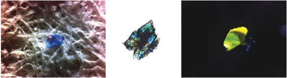 Figure 2 – Left: Impurity in paper under oblique illumination; middle: flattened paper impurity under transmitted illumination; right: paint chip under oblique illumination.
Sample manipulation is accomplished by manual adjustment of the standard microscope controls including a 1 × 3 in. travel x,y stage, coarse/fine focus, and condenser focus adjustments. By combining high-depth-of-field viewing optics, illumination options, and simple sample stage controls, the SurveyIR makes it easier to locate the sample than traditional IR microscope systems.
Being a sample compartment accessory, the SurveyIR uses the FTIR spectrometer-mounted detector. Its all-reflective optics enable data collection throughout the full frequency range of the instrument, even into the far-IR. In addition to the increased frequency range, using the onboard detector limits service and consumable cost of the liquid nitrogen-cooled mercury cadmium telluride (MCT) detector employed in traditional systems.
eSpot software from Czitek (Figure 3) allows the user to control image display, manipulation, capture, documentation, measurement, and storage. It also provides the interface for IR mode selection, illumination selection, and sample size by allowing a choice of six aperture settings down to 60 μm. Switching between different modes of spectral data collection or illumination is as simple as choosing the mode of interest from a drop-down menu at the top of the screen.
 Figure 3 – eSpot software for video microscopy live image display, capture, documentation, and analysis.
Sampling of imperfections during product manufacture
Imperfections during the manufacturing process can be costly, especially if the entire process has to be halted. SurveyIR can be implemented on the manufacturing floor to provide fast and reliable results. Depending on the size of the imperfections, they can be directly sampled via the clip-on ATR or removed and sampled on a reflective surface or an IR transparent window.
Figure 4 (left) shows a contaminant in oblique illumination that was observed in an engineered plastic molding and was excised for analysis. The excised particle was flattened and transferred to a KBr window to collect IR spectral information in transmission. Spectra on the right represent the excised contaminant (blue) and two components that showed strong correlation to the excised contaminant. The strongest correlation was that of silica gel (red), a common synthetic silicate. Ethoxylated tallow alkyl amine (purple) showed some similarity to the excised particle in Figure 4. Due to silica’s common use in the plastics industry as a filler and an anti-caking agent, it is no surprise that excised contaminant IR spectra are dominated by silica gel. The contaminant is likely a mixture of silica and natural oils such as castor or linseed oil used in the manufacturing process.
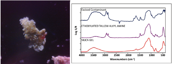 Figure 4 – Left: excised plastic contaminant; right: IR spectral interpretation of the excised particle.
Identification of contaminants in pharmaceutical production
Pharmaceutical production often uses FTIR spectroscopy to characterize raw materials and finished product. Like most manufacturing processes involving consumer goods, quality is critical. Trace contaminants are almost inevitable in large-scale production, and it is crucial to identify them quickly. Contaminants located inside the factory can cause process shutdowns and downtime, while contaminants in the hands of the consumer can damage a company’s image and reputation.
The image in Figure 5 was recorded with oblique illumination and through the diamond ATR. The spectrum of the black spot on the tablet was taken directly off the tablet without any sample preparation and is displayed as the top spectrum (blue) in the figure; the spectrum in red was collected with the diamond ATR without any sample preparation and represents the coating of the tablet. The spectrum of the black spot was searched against IR spectral databases; the most similar spectra indicated that the contaminant was comprised of ethylene–propylene copolymer, a well-known rubber material with a wide range of applications, including O-rings and gasket seals. The tablet coating is composed of hydroxypropyl methylcellulose, a common polymer coating used in controlled-release tablets. A degraded gasket that broke off during tablet processing is likely responsible for the appearance of ethylene–propylene copolymer on the tablet’s surface. With the selectivity of the ATR and the imaging quality through the diamond, the SurveyIR provides a capable microspectroscopy accessory. Features and specifications are listed in Table 1.
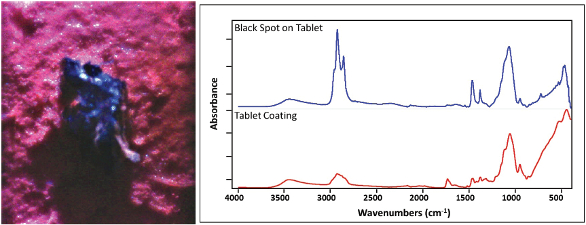 Figure 5 – Left: contaminant on surface of a pharmaceutical tablet, imaged through the diamond ATR element using oblique illumination; right: IR spectra of the contaminant and tablet coating. Analysis described in text.
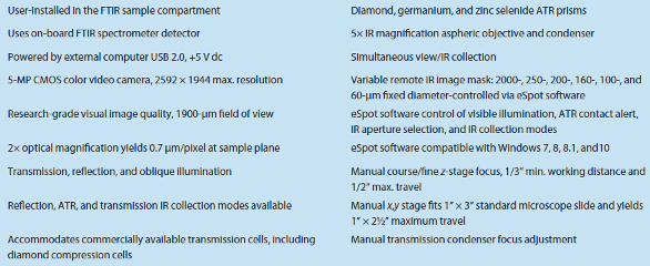
Table 1 – SurveyIR features and specifications
Conclusion
The SurveyIR is a powerful microanalysis tool that combines high analytical performance with an ergonomic, alignment-free design. Analysts can accurately characterize samples and rapidly improve quality. Additional applications include forensic science, science education, art conservation, mineral/geologic studies, drug characterization, and contaminant identification.
David W. Schiering and Anthony W. Didomenico are with Czitek, 6 Finance Dr., Danbury, CT 06810, U.S.A.; tel.: 203-456-5268; e-mail: [email protected]; www.czitek.com. John A. Seelenbinder is with PointIR Consulting, Watertown, CT.