Featured Article

Please check out our Laboratory Microscopes section for more information or to find manufacturers that sell these products.
A standard laboratory microscope
is a necessary tool in almost
every laboratory in the world.
While electron, fluorescence, confocal,
and atomic force microscopes (AFMs)
are gaining in popularity, the standard
laboratory microscope continues to be a
basic requirement. It is hard to imagine a
laboratory without one.
The standard microscope has gone through
many changes over the last few decades,
including the addition of digital imaging
via a standard trinocular configuration
with a third viewing port and various types
of automation. These include automated
slide handling, autofocus, powered zoom,
and an automated aperture. While individual
features have been automated, very few
entire procedures have been automated.
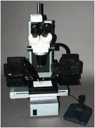
Figure 1 - The Harvester-ST.
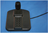
Figure 2 - The Harvester joystick.
To automate or even semiautomate many
common experiments currently performed
under a microscope would involve many
of the components mentioned above, in
addition to a patchwork of XYZ motion
control devices, manual micromanipulators,
handheld pipettors, robotic software, imaging software, temperature control stages, shakers, vortexers, etc. The SAMI
series integrated imaging system (Station
for Automated Microscopy and Imaging),
including the Harvester (FMP Products,
Inc., Greenwich, CT) automates many
standard laboratory procedures that require
use of the components mentioned above
(see Figures 1–4).
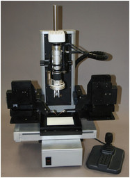
Figure 4 - The Harvester-3D.
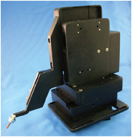
Figure 3 - System manipulator.
Automated crystal harvesting
One of the first laboratory procedures to
be automated was crystal harvesting (also
known as crystal mounting or looping a
crystal), a brief description of which follows.
The harvesting of crystals can be very
complicated. Crystals can range from several
microns to several hundred microns in
diameter. Some are almost entirely made
up of liquid (protein crystals). These tend
to be extremely fragile and are difficult
to manipulate because they often stick to
surfaces (especially certain types of plastic),
group together, move within the drop
as the user tries to harvest them, dry up
(crash out) if too much time is taken to
retrieve them from the drop, and so on.
Small-molecule crystals, such as salt, tend
to be more stable (but not always).
To begin harvesting protein or small-
molecule
crystals, a slide or plate containing
the crystals is placed under a microscope,
usually in a drop of mother liquor
(from <1 μL to greater than 10 μL) or oil,
or sometimes they are placed dry on the
microscope slide. In the case of protein
crystals, the researcher uses a small “loop”
(10–200 μm), normally made of plastic and
attached to a small handle about the size of
a pencil, and tries to manually capture the
crystal using the surface tension of the liquid
suspended within the loop. For smallmolecule crystals, the researcher can either mount the crystal in a
loop containing oil for surface tension support, or use UV-curable
glue on the end of a pin to harvest the crystal. Once the crystal is
mounted, it is flash-cooled to stabilize it and prevent it from degenerating.
At this point, the crystal is either analyzed in-house or
placed in a synchrotron—a cyclic particle accelerator in which the
magnetic field (to turn the particles so that they circulate) and the
electric field (to accelerate the particles) are carefully synchronized
with the traveling particle beam—for further investigation.
Challenges of manual harvesting
Crystal manipulation and harvesting have been done manually
since the science of crystallography began. While the looping of
larger crystals comes easily for some people, others never seem to
get the knack. Some of the problems with manually harvesting
crystals (both small and macro) include:
- Crystals are being harvested at earlier and earlier points in their
growth cycle, sometimes when they are as small as 2–5 μm. As
the crystals get smaller, the ability to manually harvest them
becomes more difficult.
- Manual harvesting may damage the crystals due to the user’s
inability to precisely control the looping tools.
- When harvesting manually,
higher levels of magnification
required for small
crystals are difficult to use
because the user’s hand
motions (i.e., shaking) are
intensified, distorting vision
and thereby affecting the
ability to accurately harvest.
- It is difficult to choose a specific
crystal to loop since
manual looping disturbs the
entire drop and any other
crystals that are present in
that drop
- Manual harvesting of crystals is done with one loop; the use of two
loops simultaneously to capture crystals is rarely done by hand.
Automated microscope platform
The basic device used was the Harvester-3D automated microscope
platform with dual micromanipulators. This robotic imaging and
sample manipulation platform offers automatic plate scanning,
3-D imaging, crystal harvesting, and a fully programmable macro
language and a complete assortment of tools. The macro language
includes commands that instruct the system to carry out its operations
unattended. Once initiated, macros can operate the microscope
for minutes, hours, or even days (i.e., time-lapse studies).
For this application, the integrated on-board options and software
included:
- A rotational microscope capable of magnifications from 50× to
400× (Hirox, River Edge, NJ)
- Autozoom and autofocus (FMP)
- A stage with automated XYZ capabilities (FMP) and multiple
axes of freedom
- Control for up to 21 motorized devices
(FMP)
- Dual multiple-axis submicron manipulators
(up to four) (FMP) on moveable
platforms
- Dual metal-halide light sources (150 W)
(Welch Allyn, Skaneateles
Falls, NY)
- Dual fiber optic pathways (simultaneous
brightfield and darkfield lighting)
- UXGA digital scientific camera
(Lumenera
Corp., Ottawa, Ontario,
Canada)
- MiTeGen ( I tha c a , NY) mount s
for looping and manipulating crystals
or the Crystal Catcher (Kyodo
International, Tokyo, Japan) (virtually
any loop or tool will work
with the system)
- Image Automation V3.65 software
for image capture and robotic control
(FMP).
System operation
Typically, the user places a coverslip or
another vessel containing the crystals
under the microscope. Because time is
usually limited (especially in the case of
protein crystals), the user presses a single
key, and the system positions the loop(s),
crystals, and microscope in preparation
for harvesting crystals. (Note: Sometimes
a small amount of Paratone-N oil
[HamptonResearch, Aliso Viejo, CA] is
used to delay the dehydration of the drop
holding the crystals to allow more time
for harvesting.)
If the user has already selected the crystal
to harvest, he/she can choose that
crystal by clicking on it with the mouse.
Once the desired crystal is selected, the
user can harvest it manually or invoke
a macro, which will harvest it automatically
based on methods and training
established by the user. Although several
off-the-shelf macros for crystal harvesting
are included with the system, the
user can program in his/her own style of
harvesting, with the system mimicking
the user’s every move. The programmable
macros control all devices available
in the automated system (XYZ stage,
micromanipulators, zoom, focus, etc.),
as well as all software functions such as image analysis, image capture, video capture,
and Z-stacked imaging.

Because the Harvester-3D can be operated
remotely, it is well suited for harvesting
oxygen-sensitive crystals within a
glove box and offers various other benefits
when used in an oxygen-free environment.
For example, with its precise joystick
operation, users do not have to work
with gloved hands, which is a tedious,
tiring, and time-consuming method of
harvesting crystals.
The micromanipulators can hold either
traditional loops (MiTeGen) or other
types of harvesting devices, such as the
Crystal Catcher. The Crystal Catcher can
be programmed to automatically harvest
both protein and small-molecule crystals
using its polymer-based adhesive technology,
and the penlike device mounts easily
onto the micromanipulators.
A typical macro for harvesting crystals is
as follows:
-
Step 1—Move stage to harvesting position.
This brings the XYZ stage into
position, autofocuses the microscope,
and moves the micromanipulators with
loops into position.
- Step 2—Mark crystal(s) for harvesting.
This highlights all of the crystals
to be harvested (either automatically
or manually).
- Step 3—Begin harvesting. Depending
on whether small- or macromolecule
crystals are being harvested,
the procedure is slightly different.
For the purposes of this discussion,
we will assume small-molecule harvesting
is being done using UV-curable
glue to attach the crystals to
the mounts.
In turn, the system will:
a) Move to the UV glue position
b) Pick up a small amount of UV glue
(amount predetermined by the user)
c) Move to the first crystal to be harvested
d) Pick up the first crystal using XYZ coordinates
generated when the user marked
the crystal (step 2)
e) Move to UV curing light for 20+ sec
to harden the glue. The user can move
the micromanipulator to neutral position
and pause so that he or she (or the
automated arm [i.e., Hitachi, Tokyo,
Japan]) can remove the harvested crystal
and replace the holder for the next
crystal to be harvested.
- Step 4—Loop to step 3 until finished
(note: macros have “if/then/else”
capabilities).
The entire process of writing the macro
to accomplish this task took about 2
hr, with a total of 10 steps. Although
the system is capable of controlling up
to 21 axes or devices simultaneously,
thi s appl icat ion requi red only 11.
Other experiments and procedures that
can be successfully programmed on the
device include autorotation and global
3-D imaging with Z-stacked images,
automatic dispensing of reagents in a
96-well plate with mixing and automated
image capture, automatic plate
scanning with 3-D rotational imaging
and image analysis, automated inspection
of parts, and autohandling and
imaging and storage.
Conclusion
With the ability to control multiple
devices, the SAMI series of automated
microscopes offers researchers a powerful
tool for designing applications or performing
specific tasks (such as harvesting
crystals) in one convenient package.
The integrated macro language allows
microscopists and researchers to create
powerful, automated solutions to imaging
and sample manipulation problems.
Please check out our Laboratory Microscopes section for more information or to find manufacturers that sell these products.
Mr. Friedlander is President, FMP Products,
Inc., 100 Melrose Ave., Ste. 206, Greenwich, CT
06830, U.S.A.; tel.: 914-939-3014; fax: 866-
226-1795; e-mail: [email protected].