Featured Article
Students learn that chromatography involves interactions of analytes with stationary phase and mobile phase, just as it was designed by Mikhail Tsvet more than a century ago. However, hydrodynamic chromatography (HDC) eludes this general definition. In HDC, particles are separated due to the different velocities of the mobile phase laminae developing across the column rather than interactions with stationary phase. In the most typical scenario, large particles are subjected to a greater velocity of the mobile phase, while smaller particles are subjected to a smaller velocity (see also the comprehensive review by Striegel and Brewer1 ). Based on this generic mechanism, a theory is available that allows one to predict the migration order and estimate the retention times of the particles with different size.
On the other hand, some separations conducted in hydrodynamic flow—also referred to as HDC—may not be exclusively based on the pure HDC mechanism. For example, interactions of particles with the surface of the capillary channel and disruption of the laminar flow by large particles may, in some cases, contribute to the separation in either positive or negative ways. Notably, HDC-like separations can be performed in macroscopic columns packed with nonporous particles or in open-tubular capillary columns (typical diameters are a few to a few tens of micrometers) without any packing material. HDC is capable of separating particles as diverse as DNA, proteins, and synthetic nanoparticles. These features make HDC a genuinely simple approach with applications spanning chemistry, biochemistry, microbiology, nanotechnology, and pharmaceutical science.
In principle, capillary HDC can be conducted using nanoflow liquid chromatography or capillary electrophoresis equipment. Such commercially available instruments enable automated and accurate injection of samples and mobile phase. Low-throughput injection can also be achieved using hydrodynamic pumping—compressing a neutral gas in the vial headspace. The flow rate of the mobile phase in the capillary column can then be estimated from the Poiseuille equation or retention times of low-molecular-weight marker analytes. The instrumentation for HDC can be custom-built using widely available items by individuals without an engineering background. Indeed, construction of HDC systems does not require extraordinary skills. However, precautions must be taken while handling compressed gases. In one such house-built system, an Arduino Pro Micro controller was used to control a pinch valve coupled with the sample/mobile phase vial.2 Thus, the sample injection time could be precisely controlled. In fact, the open-source electronic modules provide great opportunities for chemists to innovate in the area of separation science at low cost. Customized parts can readily be fabricated by 3-D printing using polymers such as acrylonitrile-butadiene-styrene (ABS) as substrates. When high gas pressures need to be applied, vulnerable parts must be fabricated of more rigid materials such as steel to match the operational specifications.
HDC can be coupled with a number of standard detectors, including light absorption, fluorescence, and mass spectrometry. Fluorescence detection is particularly useful when separating small quantities of labeled macromolecules of the same kind in thin capillaries (<5 μm). For example, labeled DNA fragments can be separated in such capillaries and detected before the outlet with the aid of a fluorescence detector.3 The high sensitivity of this class of detector is an asset because very few (in special cases, just one) molecules can produce a measurable signal. Separations in wider-bore capillaries (≥50 μm) can often be monitored by ultraviolet absorption detectors, despite their typically having over three orders of magnitude lower sensitivity. Absorbance scales with optical pathlength as well as concentration; thus, signal-to-noise ratios are smaller when using narrow-bore capillary columns than with wide-bore columns. Detectivity (limit of detection) is also affected by the baseline noise.
An HDC-like approach was recently implemented to separate large bioparticles such as liposomes and microbial and human cells.2,4 In these studies, wide-bore capillaries (e.g., 75 μm) were used as columns. Pressures as low as 100 kPa were sufficient to move the mobile phase along a 5-m-long capillary column. In fact, building and operating such “low-pressure” HDC setups is technically less demanding than building and operating higher-pressure setups for separations of small particles (e.g., DNA fragments) in very thin, long capillaries.
Although most parts of such low-pressure HDC systems are inexpensive or can be adapted from existing modular instruments, these systems can still be coupled with high-tech detectors, including mass spectrometers of various kinds as well as ultraviolet imaging detectors. As an example, electrospray ionization mass spectrometry was used to detect high-abundance lipids present in human and animal cells separated by HDC (Figure 1).4 The main purpose of the separation step here was to remove the cell medium components (salts), which would suppress ionization of the target analytes (cell membrane lipids). Interestingly, no lysis step was necessary because human and animal cells do not have cell walls and readily disintegrate in the electrospray plume.
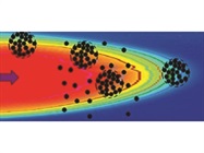
 Figure 1 – Hydrodynamic separation of liposomes and cells with ultraviolet absorption/scattering and mass spectrometric detection. (I) Schematic of the analysis setup (A). The insets show idealized principle of bioparticle separation in hydrodynamic flow (B), and disintegration of fragile bioparticles in electrospray (C). (II) Hydrodynamic chromatograms of a suspension of cells obtained by simultaneous ultraviolet absorption/scattering (blue) and mass spectrometric (black) detection. Left: mouse embryo cells (3T3-L1); right: human carcinosarcoma cells (Hs578T). Extracted ion currents (EICs) of two selected cell lipid-related species (
m/z
787, 761) and one medium-related species (m/z
413) are displayed. PLP—chromatographic feature corresponding to the cell medium components. (Adapted with permission from Ref. 4. Copyright ©2016, American Chemical Society.)In another version of this HDC separation setup,2 an ultraviolet imaging detector incorporating an active pixel sensor was used (ActiPix D100, Paraytec Ltd., York, U.K.). With this kind of detector, multiplexed separations can easily be performed. The multiplexing capability is important when fingerprinting suspensions of large bioparticles. Such mixtures tend to sediment. Despite the samples being shaken prior to the analyses, noticeable vertical gradients developed along the sample vials within a few minutes. As a consequence, if the HDC runs were not performed immediately, the proportion of large cells and cell aggregates was greater near the bottom than at the top of the vial during the injection step. The bioparticle gradient compromises the sample homogeneity that would normally be taken for granted. However, one could turn the sedimentation process to advantage, and use it for a partial preseparation of complex bioparticle mixtures prior to injection of sample to the separation column. For example, in one of the variants, three parallel capillary columns were implemented. The inlets of these columns were positioned at different heights within the sample vial: One of them sampled the suspension near the bottom of the vial, the second sampled the middle, and the third the top. As expected, sedimentation time before the HDC separation had an effect on the chromatograms; longer sedimentation times contributed to more pronounced vertical gradients in the vial.2 While the above example illustrates the possibility to detect bioparticles present in different regions of heterogeneous samples, multiplexed HDC can also be utilized in other ways, for example, to analyze multiple samples at the same time.
Hydrodynamic flow exerted in microscale capillaries can be utilized to perform other types of measurements beyond separation. In one recent study, an instrument with UV imaging detection was used to characterize aggregates of cyclodextrin–drug complexes (Figure 2A).5 In this case, partial separation of aggregated and nonaggregated cyclodextrin–drug complexes was achieved (Figure 2B). Fitting the features related to the dispersive and convective components enabled deconvolution of the recorded “Taylorgrams” and determination of the proportions of the aggregated form. This is possible since UV detection gives mass-weighted response, unlike dynamic light scattering, where the response strongly favors the aggregate. The apparatus used in this study (Viscosizer 200, Malvern PANalytical [formerly Malvern Instruments Ltd.], Malvern, U.K.) has two windows on the capillary (ID, 75 µm; total length, 130 cm). Measurement of peak broadening in flow between the two and fitting using Taylor dispersion analysis (TDA) provides information on diffusivity and hydrodynamic size of the nonaggregated form.
 Figure 2 – Characterization of aggregates of cyclodextrin–drug complexes in hydrodynamic flow. (A) Solvent velocity distribution across capillary; (B) sizing records at 280 nm and data fitting—primary data (gray) and fit (black) using model with dispersive (dashed) and convective (dotted) components. (Adapted with permission from Ref. 5. Copyright ©2017, Elsevier.)
In TDA measurements on heat-stressed bovine serum albumin (BSA) samples, small proportions of aggregates were observed as pre-peaks before the main BSA band.6 With UV imaging detection at 214 nm, individual protein particles with size > ~1 μm separated by HDC can be identified as sharp spikes that travel at constant velocity between the two windows.
Conclusion
Hydrodynamic chromatography has many potential uses in the food industry, where complex functional food matrices such as yogurts can be analyzed directly after simple dilution. The ability to use HDC to reveal dynamics of the matrix microbiome during primary fermentation and monitor the growth of large aggregates could also have relevance in the biotherapeutics sector.
Although the future of hydrodynamic separations and sizing of particles is promising, hydrodynamic separations need to be further popularized. For example, the topic can be incorporated to the syllabi of undergraduate and graduate courses related to analytical science. It is useful to start with the basic concepts of fluid dynamics, characteristics of laminar flow, and diffusion-driven phenomena. Then, one can discuss examples of the techniques that take advantage of hydrodynamic properties of microscopic particles to separate them and/or to determine their size, including HDC, TDA, as well as field flow fractionation, split flow thin cell fractionation, and ultracentrifugation. Understanding the principles of those convenient analytical techniques is certainly useful for trainees interested in the classical branches of science as well as newer directions such as nano- and microtechnology.
References
- Striegel, A.M. and Brewer, A.K. Hydrodynamic chromatography. Ann. Rev. Anal. Chem.2012; doi: 10.1146/annurev-anchem-062011-143107.
- Tang, Y.-R.; Huang, H.-Y. et al. Capillary hydrodynamic chromatography reveals temporal profiles of cell aggregates. Anal. Chim. Acta2016; doi: 10.1016/j.aca.2015.12.049.
- Friedrich, S.M.; Liu, K.J. et al. Single molecule hydrodynamic separation allows sensitive and quantitative analysis of DNA conformation and binding interactions in free solution. J. Am. Chem. Soc.2016; doi: 10.1021/jacs.5b10983.
- Chen, S.-Y.; Wu, C.-Y. et al. One-step detection of major lipid components in sub-microliter volumes of unpurified liposome and cell suspensions. Anal. Chem.2016; doi: 10.1021/acs.analchem.6b01740.
- Zaman, H.; Bright, A.G. et al. Characterisation of aggregates of cyclodextrin-drug complexes using Taylor dispersion analysis. Int. J. Pharm.2017; doi: 10.1016/j.ijpharm.2017.02.012.
- Hulse, W.L.; Gray, J. et al. Evaluating the inter and intra batch variability of protein aggregation behaviour using Taylor dispersion analysis and dynamic light scattering. Int. J. Pharm. 2013; doi:10.1016/j. ijpharm.2013.05.062.
Pawel L. Urban is with the Department of Applied Chemistry, National Chiao Tung University, 1001 University Rd., Hsinchu, 300, Taiwan; e-mail: [email protected]; www.nctu.edu.tw/en. David M. Goodall is with Paraytec Ltd., York, U.K. P.L. Urban acknowledges the Ministry of Science and Technology, Taiwan (MOST 104-2628-M-009-003-MY4), for financial support.