Featured Article
Multiphoton microscopy is a powerful research tool that combines laser scanning microscopy with long wavelength multiphoton excitation to capture high-resolution three-dimensional images of biological specimens. Contrast can come from a variety of sources, ranging from introduced fluorescent dyes and proteins to intrinsic biomolecules with inherent fluorescent or nonlinear scattering responses.
This methodology is particularly useful to researchers seeking to study dynamic processes in living cells and tissues without inflicting significant damage upon or disruption to the specimen. Multiphoton microscopy can access sites several hundred micrometers below the surface without having to physically cut into the sample. The longer wavelengths used in multiphoton microscopy also translate to reduced phototoxicity, enabling observation over longer durations.
Comparing specialized microscopy techniques
Traditional widefield fluorescence microscopy can provide submicron resolution of biochemical events in living systems. However, the technique is limited in sensitivity and spatial resolution by background noise caused by secondary fluorescence from regions above and below the focal plane. Confocal microscopy circumvents this problem by rejecting out-of-focus background fluorescence with pinhole apertures to produce thin (less than a micron) unblurred optical sections from within thick specimens. However, penetration, especially in live tissue specimens, is typically limited to around 100 micrometers.
Multiphoton microscopy provides an alternative to confocal techniques through selective excitation of just the focal plane, thus eliminating out-of-focus fluorescence at the onset. In the past, the high cost and complexity of multiphoton microscopy systems limited use of this technique. More recently, however, increasingly easier-to-use multiphoton systems have made the benefits of multiphoton microscopy more accessible for many investigations.
The principal benefits of multiphoton microscopy can be summarized as follows:
- Detail: high-resolution 3-D imaging with a range of intrinsic and extrinsic contrast labels
- Dynamics: high-speed and/or longitudinal imaging of live cells and tissue under in vivo and in vitro settings
- Depth: deep imaging through intact highly scattering tissue.
Basic principles of multiphoton microscopy
Multiphoton excitation occurs when multiple photons simultaneously interact with a molecule to emit a single shorter-wavelength photon.
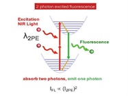
The most common mechanism of interaction is fluorescence, such as in two-photon excited fluorescence (Figure 1), but other optical processes such as nonlinear scattering (e.g., second harmonic generation) or coherent Raman scattering can also be utilized. The key element is that these multiphoton interactions require very high excitation intensity. In fact, ultrashort pulsed infrared lasers and high numerical aperture objective lenses are critical to achieving the necessary density of photons in time and space. The resulting multiphoton excitation is then strongly localized to the focal volume of the objective.
 Figure 1 – Two-photon excited fluorescence is a common contrast mechanism employed in multiphoton microscopy.
The compact excitation volume of a multiphoton microscope enables optical sectioning within thick biological specimens without the need of a confocal pinhole aperture. By eliminating the pinhole, more emission photons can be collected and the signal-to-noise ratio is much improved. This is especially true when imaging sites deep within highly scattering samples. Deep penetration in biological tissue, typically several hundred micrometers below the surface, is further facilitated by the use of near-infrared excitation wavelengths that experience less scattering than visible light when traveling through the turbid material.
 Figure 2 – Sensory cortex of anesthetized Thy1-YFP-H mouse. Acquired at the RIKEN Brain Science Institute, Olympus Collaboration Center. (Courtesy of Dr. Hiromu Monai, Dr. Hajime Hirase, and Dr. Atsushi Miyawaki.)
Additional benefits of multiphoton microscopy include reduced photobleaching due to the highly localized excitation. Phototoxicity in live cells is also reduced by the shift in excitation from the ultraviolet to the near-infrared range. All these advantages enable researchers to conduct experiments on thick living tissue under both in vitro and in vivo environments. Under the right conditions, images can be captured from sites over 1 mm deep in intact tissue with subcellular resolution—a feat difficult, if not impossible, with other microscopy techniques (Figure 2).
Optimizing deep imaging performance
It is worth noting that optical resolution in multiphoton microscopy does not exceed that achieved with confocal microscopy. The real benefit lies in its ability to capture images from deeper below the surface than other high-resolution techniques. So how can we optimize deep imaging performance in a multiphoton microscope to fully leverage this advantage? Most methods for improving microscope performance comprise two fundamental actions: increasing excitation and improving detection efficiency.
Recall that multiphoton excitation is dependent on achieving very high intensity in a small volume. The challenge with deep imaging is to maintain this intensity when focusing further below the sample surface. Here are some approaches:
- Scaling laser power with depth. Increasing laser power is the simplest way to increase excitation intensity. However, laser power is typically limited by tissue damage at shallow sites. Using today’s technology, software-controlled optical modulators can scale laser power with z-position such that more power is delivered at deeper sites. This helps compensate for scattering and absorption losses as the laser travels through the tissue.
- Adjusting the correction collar. Refractive index mismatch causes spherical aberration and degrades the focus. These aberrations progressively worsen with depth, stretching the focal volume and decreasing the excitation intensity. An objective correction collar can compensate for spherical aberration to preserve both resolution and contrast. Ideally, collar settings should be optimized at each z-position. This can be facilitated by a motorized correction collar system and can even be automatically driven based on measured image contrast.
- Underfilling the objective. In highly turbid specimens, scattering can be the larger cause of focus degradation. While matching the laser beam diameter to the back aperture of the objective lens typically ensures maximum resolution, reducing the input beam diameter allows more laser power to reach the focal region. Underfilling the objective compromises image resolution, but a system with a continuously tunable beam expander allows the desired tradeoff balance.
Inherent optical sectioning means that multiphoton microscopy is about collecting as much light as possible. Every detected photon is assumed to originate from the focal volume and contribute to useful signal. Strategies then involve improving detection and collection efficiency.
- High-sensitivity GaAsP detectors. For deep imaging, high-sensitivity gallium arsenide phosphide (GaAsP) detectors are advantageous. The quantum efficiencies of these detectors are among the highest available for visible wavelength ranges.
- Large FOV objective. The photodetector is only one component of the detection system. The first, and arguably most important, element is the objective. Low-magnification objectives capture more of the scattered emission because of a larger field of view (FOV). Users should be mindful of vignetting, which becomes an issue, both within the objective and along the path to the non-descanned detectors. Scattered photons tend to travel at larger angles relative to the optical axis of the objective. Objective magnification does not always tell the full story for field of view. An objective designed for multiphoton microscopy should also be designed for low vignetting.
Conclusion
Multiphoton microscopy has become a method of choice for dynamic high-resolution imaging of living cells in thick tissues and live animals. The technique is also useful in biological systems where ultraviolet excitation would not otherwise be possible due to the light transmission characteristics or photochemical impact on the specimen. Side effects such as photobleaching and photodamage are minimized in multiphoton excitation, and occur only in the immediate focal volume. Continued trends in the development of longer-wavelength pulsed laser sources, red-shifted fluorophores, and higher-order imaging modalities such as three-photon excited fluorescence promise to deliver even deeper imaging than currently demonstrated. Meanwhile, advances in tighter integration and automation of multiphoton microscope systems make the technique more accessible to a wider audience of researchers.
Carlo Amadeo C. Alonzo is associate product manager, Multiphoton Microscopy, Olympus Corporation of the Americas, Scientific Solutions Group, 48 Woerd Ave., Waltham, MA 02453, U.S.A.; 781-419-4855; [email protected]; www.olympus-lifescience.com