Featured Article
Cytometry captures more data than ever and across multiple formats
To study a wide range of particles in liquids, scientists use flow cytometry. The sample can be tagged with fluorescent markers that light up as the particles pass a laser in the device. Other methods can be incorporated. A flow cytometer can be used to detect specific particles or cells, sort them, and more.
At the University of Cádiz in Spain, doctoral researcher in marine and coastal management Esther Bautista Chamizo and her colleagues used flow cytometry to study the marine diatom Phaeodactylum tricornutum. 1 The scientists wanted to see how climate change—and the possible impacts on pH, temperature, and salinity—could affect microalgae. Chamizo and her colleagues used flow cytometry to measure growth, cell viability, metabolic activity, and cell properties, including size and autofluorescence. The researchers noted: “The results showed adverse effects on cultures exposed to pH6 and high temperature and salinity.”
For a scientist interested in using flow cytometry, many platforms can be considered. Moreover, many features also come into a purchase decision. Developed in 1968, flow cytometry celebrates its 50th birthday this year, and new products keep hitting the market.
 Flow cytometry can be used in many ways. Here, smooth muscle cells are being examined for use in tissue engineering at the Georgia Institute of Technology Emory Center. (Image courtesy of the Georgia Institute of Technology Emory Center.)
 Ongoing improvements
Despite half a century of development, there is no sign of flow-cytometry platforms slowing down in innovations. In September 2017, Beckman Coulter (Indianapolis, IN) revealed some of the advances underway when it released the CytoFLEX LX, which covers a wide range of excitation wavelengths, from the near ultraviolet (375 nm) to infrared (808 nm). For collecting information, it uses avalanche photodiode detectors that, the company states, provide “high and stable quantum efficiency from 400 to 1100 nm.”
Despite providing so much selection, this platform just sits on a benchtop—not taking up a lot of room in a lab. And scientists get a range of options in selecting the right platform, and then quickly using it. As the company states: “Researchers can unpack, set up, and run an experiment within an hour—and select from three models and 47 configurations of the CytoFLEX benchtop research analyzer series, which sets new standards for fluorescence sensitivity.”
Plus, putting flow cytometry on a benchtop can be done with various platforms. For example, the NovoCyte Quanteon (launched in May by ACEA Biosciences, San Diego, CA) sits on a bench, but small size doesn’t mean low power. As the company notes: “The NovoCyte Quanteon employs a series of silicon photomultipliers (SiPM), which have superb detection sensitivity. This high-performance instrument is capable of detecting up to 27 parameters with enhanced resolution and walk-away automation capabilities.” Plus, it works with samples in many formats, including single tubes; multitube racks; or 24-, 48-, 96-, or 384-well plates.
Other manufacturers also provide flow-cytometry platforms with broad capabilities. As an example, the 2100 Bioanalyzer from Agilent (Santa Clara, CA) can be used with DNA, RNA, or proteins to measure size and quantify and determine purity of a sample. Like many modern flow-cytometry platforms, the 2100s are easy to use. As Agilent says: “load the chip, press ‘start,’ and the instrument does the rest!” Making it even easier, Agilent offers ready-to-use assays and reagent kits for the 2100s. Plus, it only takes 1–4 microliters of sample to run an assay.
These examples already give some idea of what the market offers, and—more important—what scientists should expect. That can include a wide range of excitation wavelengths, equally wide-ranging detectors that are accurate and sensitive, plus ease-of-use features that range from validated assays and reagents to simple operation and analysis.
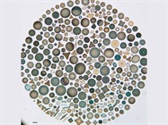
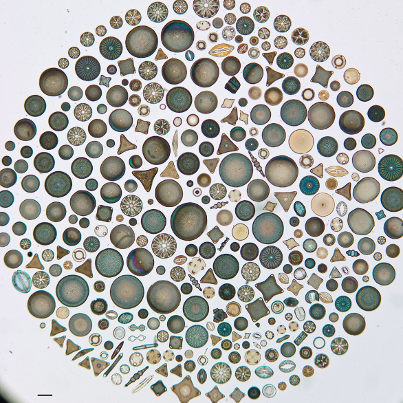 Scientists use flow cytometers to analyze cells and particles, and even the impact of climate change on marine phytoplankton, such as the diatoms shown here. (Image courtesy of the California Academy of Sciences.)
Adding options
One option that some scientists want with flow cytometry is imaging, which is like combining the capabilities of traditional flow cytometry with a microscope. Then, for example, the flow cytometry can be used to isolate specific cells or particles, which can then be analyzed visually.
From San Jose, California, Molecular Devices offers the SpectraMax MiniMax 300 Imaging Cytometer. According to the company, this platform, “enables quick imaging and analysis of cells and gives you a front-row view of phenotypic changes that accompany cytotoxicity, cell proliferation, and protein expression.”
Even better, the imaging capability is an upgrade option for the company’s SpectraMax i3/i3x Multi-Mode Detection Platform. This upgrade can even be performed in the field, rather than sending a flow cytometer back to the manufacturer.
One of the main advantages of adding imaging is the resulting increase in data. Instead of just counting, sorting, or measuring cells, scientists can look at them. That opens the door for many more analytical options, especially with the platform’s SoftMax Pro Microplate Data Acquisition & Analysis Software.
In most flow-cytometry experiments, scientists use one or more fluorophores to label cells or targets. That’s easy enough with one fluorophore, but not so easy with a few. So, Thermo Fisher Scientific (Waltham, MA) created its Fluorescence SpectraViewer. With this online tool, scientists can test the compatibility of combining specific fluorophores. That information can be used in designing an experiment, rather than finding out afterward that the fluorophore signals overlapped too much for accurate measurements or detection.
That’s not the only technology that Thermo Fisher suggests combining with flow cytometry. Where automation really matters, the company can put together a collection of platforms to create an advanced workflow. For example, the company’s Invitrogen Attune NxT Flow Cytometer can be combined with the Invitrogen Attune NxT Autosampler, Invitrogen External Fluid Supply, and Thermo Scientific Orbitor RS 2 Microplate Mover. The company calls this setup a “comprehensive multicomponent automated workcell.”
The most important “option” in flow cytometers is the expanding selection that scientists can consider. In April 2018, for instance, MilliporeSigma—the Life Science business of Merck KGaA, Darmstadt, Germany—unveiled its CellStream flow cytometry system, which uses a camera for detection. It can use as many as seven lasers to analyze cells and submicron particles.
So, even at 50 years old, the flow-cytometry market seems poised to speed up, not slow down. It’s not just the improvements in excitation and detection options that matter, but also the improvements in add-on features, such as imaging and automation. As the ability to collect larger volumes and different types of data increases, flow cytometers will give scientists more options for exploring “big data” and the correlations between it. As a result, the bioinformatics side must keep pace with the hardware advances, because there’s no slowing in today’s flowing—not when it comes to analyzing cells and particles sweeping by advanced detectors.
Reference
- Bautista-Chamizo, E.; et al. Will temperature and salinity changes exacerbate the effects of seawater acidification on the marine microalga Phaeodactylum tricornutum? Sci. Total Environ. 2018; doi: 10.1016/j.scitotenv.2018.03.314 [epub ahead of print].
Mike May is a freelance writer and editor living in Texas. He can be reached at [email protected]