Featured Article
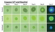
Image-based, automated analysis of 3-D tumor spheroids in microwell plates is becoming standard practice for the evaluation of anticancer drug compounds. Image cytometers are ideal for the rapid analysis and characterization of 3-D tumor spheroids, providing both quantitative and qualitative data.
The Celigo benchtop image cytometer (Nexcelom Bioscience, Lawrence, MA) is capable of reading microwell plates, among other cell-culture vessel formats. Its use of brightfield and fluorescent imaging enables high-speed, fully automated imaging and quantification of suspension and adherent cells, as well as many other cell models such as tumor spheroids.
Tumor spheroids in drug-induced/growth inhibition tumor studies are rapidly characterized and quantified based on size, migration and invasion,1 viability, dose response, and monitoring a single cell to a single spheroid formation.2 An example of data obtained for a drug treatment, kinetic time-course study is shown in Figure 1.
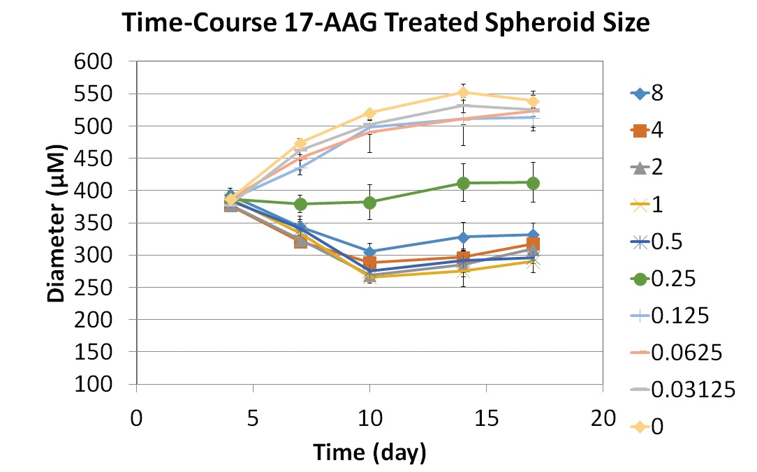 Figure 1 – Time-course study of 17-AAG effect on spheroid size. Kinetic time-course experiments can be carried out to measure tumor spheroid size as it relates to the dose-dependent drug treatment. The experiment shown was performed over a 17-day period to assess the efficacy of 17-AAG on U87MG spheroids.2
Celigo’s proprietary optics and scanning enable fast imaging of the entire well in brightfield and four fluorescence channels (green, red, far-red, and violet). The combination of brightfield and fluorescent imaging allows for the development of multicolor assays for both 2-D and 3-D assays (see Figure 2). Researchers need not stop an experiment after completing the drug treatment course on day 17—they can gather more data by performing a fluorescence-based viability or apoptosis assay to determine whether the drug of interest inhibits spheroid growth or is cytotoxic (see Figure 3).
 Figure 2 – Tumor spheroid viability measurement. U87MG spheroids were stained with Calcein AM (green) and PI (red) to determine spheroid viability. The control tumor spheroid increased in size over time and shows an increasing necrotic center with the PI staining, while the 17-AAG treated spheroid not only decreased in size but also is seen to fragment into pieces at later time points.2
The Celigo image cytometer makes it possible for researchers to gain a deeper understanding of how drugs may affect the tumors under study. It can also be employed for high-throughput screening experiments of cancer drug candidates. The system allows users to eliminate manual microscopy, replace end-point assays with kinetic assays, and increase the efficiency and speed of data collection.4
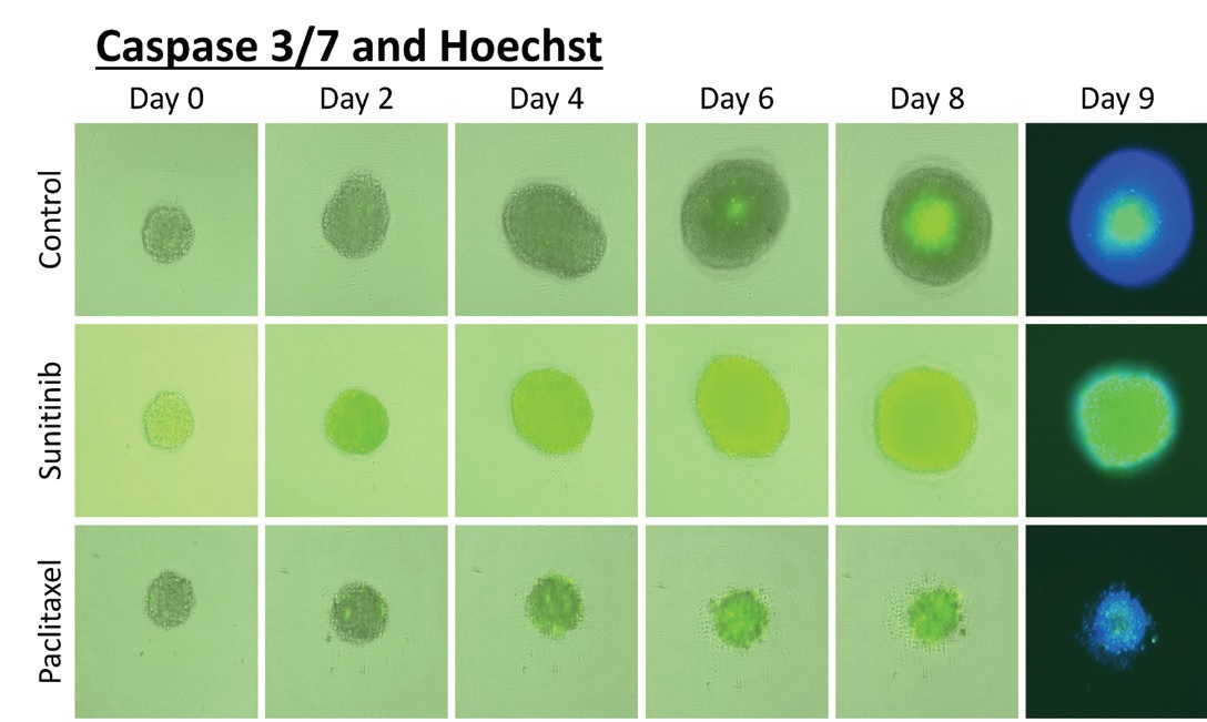 Figure 3 – Tumor spheroid apoptosis measurement. Time course of brightfield and fluorescent images were captured and overlaid to examine apoptosis (caspase 3/7) in U87MG tumor spheroids under different conditions. Control tumor spheroids increased in size over time and show an apoptotic core at days 8 and 9. The kinetic effects indicated by sunitinib malate (apoptotic), paclitaxel (late cytotoxic) at 5 μ
M
show very different drug effects. Spheroids treated with sunitinib increase in size over time and have a very high level of apoptosis, while spheroids treated with paclitaxel do not grow in size and are not very apoptotic.3References
- Cribbes, S.; Kessel, S. et al. A novel multiparametric drug-scoring method for high-throughput screening of 3D multicellular tumor spheroids using the Celigo image cytometer. SLAS Discov. 2017, 22(5), 547–57.
- Kessel, S.; Cribbes, S. et al. High-throughput 3D tumor spheroid screen- ing method for cancer drug discovery using Celigo image cytometry. SLAS Technol. 2017, 22(4), 454–65.
- Kessel, S.; Cribbes, S. et al. Real-time apoptosis and viability high- throughput screening of 3D multicellular tumor spheroids using the Celigo image cytometer. SLAS Discov. 2018, 23(2), 202–10.
Dmitry Kuksin, Ph.D., is product manager, Nexcelom Bioscience, 360 Merrimack St., Lawrence, MA 01843, U.S.A.; tel.: 978-327-5340; e-mail: [email protected]; www.nexcelom.com