Featured Article
Protein adsorption, polymer-based adhesion, and protein chromatography are closely related but separate scientific disciplines. Under gradient chromatography conditions, most proteins (or biopolymers) are adsorbed to the chromatography support until the solvent has changed sufficiently to encourage desorption and elution. The period of time that the biopolymer is bound to the support material provides sufficient conditions for adhesion. All three of these disciplines (adsorption, adhesion, and chromatography) follow the same physical binding processes.
The authors examined protein adhesion to a number of HPLC supports in gradient chromatography and noted substantial loss of material when either weak ion exchange or hydrophobic interaction HPLC was used.1,2 More extensive losses using gradient elution in reversed-phase HPLC have also been reported.3 Losses are dependent on the retention time, temperature, and molecular weight of the proteins. There may be other important factors such as pH and eluent composition, but these have not been examined fully for protein losses in chromatographic separations.
In general, increased exposure time to the sorbent, elevated temperatures, and larger molecular weights increase protein loss.4-6 Isocratic separations are less susceptible to permanent loss because the biomolecules are not held on the sorbent for prolonged periods. Thus, caution must be practiced when gradient-based HPLC separation is applied when quantifying the amount of material present or for accurate identification of all components of a complex sample. Some components may be so strongly retained by the media that they will not elute under normal operating conditions.7 The long-term or permanent retention of these components can also influence future separations as the chemistry of a portion of the column surface will be modified by the adsorbed substances.
HPLC supports are designed to maximize the sorbent surface area and hence optimize the resolution of the separation. Therefore, the separation itself can be considered investigative into the protein adsorption process. Protein adsorption8,9 and polymer adhesion10 have been studied extensively. Both typically involve the study of polymers (or biopolymers) at synthetic surfaces. In adhesion, it has been shown that binding becomes stronger with time4 and is more rapid for higher-MW polymers.5,6
The current authors showed the same trend for proteins when adsorbed to ion exchange1 and hydrophobic interaction substrates in HPLC separations.2 Losses in reversed-phase HPLC have also been reported.11 The relationship between MW and losses appears to be logarithmically related to the molecular weight of the species when a variety of retention factors are considered and can be generalized as:
L = m(log MW) + B
Where L is the loss of the biopolymer or protein, and m is the slope of the straight line that depends on the time of retention, surface chemistry, and temperature. The slope is also dependent on the log of the molecular weight. The Y-intercept, B, is almost always below the origin because HPLC sorbents are designed to be less adhesive to biopolymers, that is, low-MW components are sorbed reversibly to the column material. An example of this correlation can be seen in Figure 1, where several proteins were investigated separately under identical conditions. These proteins were held on an ion exchange support for varying periods of time to examine their rate of loss. All the proteins except myoglobin followed the log MW versus loss linear relationship. The accelerated loss of myoglobin was unexpected. The intercept, B, is typically negative for HPLC supports, indicating that low-MW substances are not bound to the
substrate. Both the slope and intercept are well worth exploring, especially by those who produce column support materials to help minimize losses. Of all the soluble proteins studied, only myoglobin deviated significantly from this straight line when examined under identical column (or surface) chemistries and temperature.12
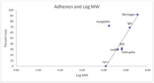 Figure 1 – This graph shows the % loss and log MW for seven proteins. Beta galactosidase is a homodimer consisting of two subunits of approximately 65 KDa. Separations were performed at 37 °C on an anion exchange column. Losses were calculated after being held on the column support for 9 minutes. (Image reproduced with permission from the authors and Ref. 12.)
The reasons for the similar behavior of several proteins are likely related to the similarities in the surface chemistry of the proteins. When in aqueous solution, the most hydrophilic side chains are generally exposed, making them appear similar to one another when compared to the synthetic substrate or sorbent.13 Deviations from this linear trend may be explained through unusual externally exposed moieties or other peculiar structural features.
Simple geometric relationships between the sorbent surface and protein may be used to model and help confirm the log MW relationship described above if it is assumed that all proteins are spherical and the region of adhesion spans a finite distance from the flat sorbent. Certainly not all proteins are spherical; however, the model presents a reasonable first-order approximation of the adsorption process due to the surprisingly linear behavior of so many proteins.12
These losses occur over minutes rather than microseconds. For example, one might lose 50% of a fibrinogen sample (MW ca. 340,000) in 1 minute during a linear gradient depending on the column chemistry and temperature.1 In contrast, the much smaller protein, cytochrome c (MW ca. 12,000), was typically fully recovered under similar conditions.14 Therefore, faster gradients can help minimize losses.
To help monitor losses when unknown samples are analyzed by gradient HPLC, one should perform a mass balance simply by placing a low-dead-volume connector in place of the column and injecting a portion of the sample to identify a 100% recovery peak area. Most detector/integrator combinations will give accurate recovery data in this manner when operating in their linear range. If 100% recovery is not obtained when the sample is then eluted through the column, an analysis using SDS polyacrylamide gel electrophoresis or gel permeation chromatography will help determine if larger (>ca. 15 KDa) MW biopolymers are in the sample and have been lost in the media.
Conclusion
Gradient elution of biopolymers by HPLC is an effective method for separation and quantitation, but when higher-MW biopolymers are analyzed, one should check the recovery to determine whether the peak areas are accurate. When unknown samples are analyzed, there is a chance that some of the sample has been lost, and there are ways to determine that without procuring expensive additional equipment. Finally, since sample components can be lost, frequent column cleaning is a worthwhile endeavor. The authors have used 0.1–0.01% SDS in dilute buffer to routinely clean columns and that is somewhat effective. However, since adhesion is a time-dependent process, cleaning should be carried out immediately following each separation, followed by rinsing to remove residual SDS.
These findings also impact the behavior of biopolymers in living systems. Additional work needs to be done to determine how adhesion is involved in biopolymer function in vivo, and also to verify similar losses in other biopolymers such as plasmids, glycoproteins, polycarbonates, etc., when gradient chromatography is used or surface adhesion is involved in processing.
References
- Goheen, S.C. and Hilsenbeck, J.L. High-performance ion-exchange chromatography and adsorption of plasma proteins. J. Chromatogr. A 1998, 816, 89–96.
- Goheen, S.C. and Gibbons, B.M. Protein losses in ion exchange and hydrophobic interaction HPLC. J. Chromatogr. A 2000, 890, 73–80.
- Stadalius, M.A.; Gold, H.S. et al. Optimization model for the gradient elution separation of peptide mixtures by reversed-phase high performance liquid chromatography: verification of retention relationships. J. Chromatogr. 1984, 296, 31–59.
- Plunkett, M.A. and Rutland, M.W. Dynamic adhesion of grafter polymer surfaces as studied by surface force measurements. J. Adhesion Sci. Technol. 2012, 16(7), 983–96.
- Kajtna, J.; Golob, J. et al. The effect of polymer molecular weight and crosslinking reactions on the adhesion properties of microsphere water-based acrylic pressure-sensitive adhesives. Int. J. Adhesion and Adhesives 2009, 29(2), 186–94.
- Galliano, A.; Bistac, S. et al. Adhesion and friction of PDMS networks: molecular weight effects. J. Colloid Interface Sci. 2003, 265(2), 372–9.
- Bobaly, B.; Sipko, E. et al. Challenges in liquid chromatographic characterization of proteins. J. Chromatogr. B 2016, 1032, 3–22.
- Andrade, J.D. and Hlady, V. Protein adsorption and materials biocompatibility: a tutorial review and suggested hypotheses. Adv. in Polym. Sci. 2005, 79, 1–63.
- Roach, P.; Farrar, D. et al. Interpretation of protein adsorption: surface-induced conformational changes. J. Amer. Chem. Soc. 2005, 27(22), 8168–73.
- Zhao, Y.-P.; Wang, L.S. et al. Mechanics of adhesion in MEMS—a review. J. Adhes. Sci. Technol. 2012, 17(4), 519–46.
- Pearson, J.D. High-performance liquid chromatography column length designed for submicrogram scale protein isolation. Anal. Biochem. 1986, 152(1), 189–98.
- Patananan, A. and Goheen, S.C. The surface-mediated unfolding kinetics of globular protein is dependent on molecular weight and temperature. J. Undergrad. Res. 2008, 8, 97–104.
- Rose, G.D.; Geselowitz, A.R. et al. Hydrophobicity of amino acid residues in globular proteins. Science 1985, 229, 834–8.
- Herbold, C.W.; Miller, J.H. et al. Cytochrome c unfolding on an anionic surface. J. Chromatrogr. A 1999, 893, 137–46.
Steven C. Goheen is retired from Pacific Northwest National Laboratory, Richland, WA, U.S.A. Jacqueline Hilsenbeck-Fajardo is with the University of Delaware, 210 S. College Ave., Newark, DE 19716, U.S.A.; e-mail: [email protected]; www.udel.edu. The financial assistance of Pacific Northwest National Laboratory and the U.S. Department of Energy is gratefully acknowledged. The authors are grateful for the assistance of many students and staff. Contributors included Craig Herbold, Betty Gibbons, and Alexander Patananan.