Featured Article
Olympus was founded by Japanese businessman Takeshi Yamashita in 1919 with the mission “to create something truly original and bring new value to society.” It is this spirit of creation that launched the vision of Olympus, which originally specialized in microscopes and thermometers.
There have been many milestones in its nearly 100-year history that have shaped Olympus, but it is the developments in the modern era that have most notably impacted life science research as we know it today. Each evolution in technology has offered subsequent advances in the imaging solutions and techniques that researchers have available to them. For instance, the 1970s were marked by a diversification of microscopy needs. To meet the demands of these industry-wide developments, Olympus embarked on providing upright microscopes categorized by the applications they served, including research (AH Series), clinical laboratory (BH Series), and education (CH Series) (see Figure 1).
 Figure 1 – From left: AH Series, BH Series, and CH Series.
The company’s flagship microscope at the time was the VANOX AH, and it was the first microscope to use a “platform”-based design. The BH series was the first step in creating a universal frame for multipurpose use. BH microscopes could be transformed to meet various specifications, including polarization, phase contrast, differential interference, and simple transmission fluorescence microscopy. By merely switching the microscope head or lens modules, users could vary the observation method. The CH series was an educational microscope for teaching purposes in biological research and the clinical laboratory field. Because of its modular design, the microscope could be used for simple polarization, drawing, or epi-illumination (metallurgical) microscopy.
Function meets technology
In 1983, the VANOX AH2 became the first microscope implemented with an autofocus mechanism, which was later applied to the autofocus function for single-lens reflex cameras. This microscopy advancement was rooted in the use of a motorized device used to automatically set the field stop, aperture stop, and condenser selection, in line with the objective lens specifications (magnification and brightness). The use of cutting-edge electronics resulted in an easy-to-operate microscope that enabled users to concentrate on observation.
Also developed during this period, the BH2 series was the starting point of configurable microscope systems. One feature of this microscope was its long barrel (LB) objective lens series (1×–100× oil) that could be used for brightfield, polarization, fluorescence, and phase contrast microscopy. This led to the launch of the SZH high-end stereomicroscope.
Developments leading to today’s microscopy
In the life sciences, advances in fluorescent protein labeling techniques in the 1980s and ’90s triggered the need for more automated systems to automatically record the rainbow of colors now being used to identify cellular structures. This trend converged with the modification of green fluorescent protein (GFP) for live cell imaging, which became one of the most important advances in the life sciences—influencing the methods used to view dynamic processes inside living cells that were previously invisible, such as nerve cell development in the brain and the spread of cancer cells. This led to the demand for live cell imaging techniques and higher-sensitivity, lower-phototoxicity detection technologies.
In the 1990s, Olympus introduced UIS (universal infinity system) optics with dramatically improved image quality and an infinity optical system that allowed users to customize microscopes with a variety of optical components. These optics were used in the AX/BX/CX series, which offered outstanding image quality and an ergonomic design.
During this time the computer revolution started to emerge and was leading to greater automation across all industries. Microprocessors, CPUs, GPUs, and memory were evolving to meet some of the most difficult challenges in modern science. To support high-end research and daily laboratory work, Olympus introduced FLUOVIEW microscopes and the DP camera series. FLUOVIEW laser scanning confocal microscopes selectively acquire sectioned layers of the sample, scanning with an excitation laser and producing a 3-D image (Figure 2). The DP series are microscopy-dedicated digital cameras for research and recording.
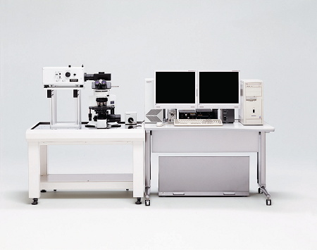 Figure 2 – FV300 confocal laser scanning microscope.
Improvements in image quality and automation
In 2003, Olympus launched the FLUOVIEW FV1000 laser scanning microscope, which was implemented with twin-laser scanners: one for imaging and the other for stimulation. This combination significantly improved sensitivity, and simultaneous stimulation provided the opportunity for real-time imaging during photostimulation. The multiphoton model of the FV1000 microscope was launched three years later. Multiphoton laser scanning microscopes could excite only the fluorescence molecule in an optically focused region without exciting other areas (which leads to background noise). They also provided the capability to deep-dive into brain samples with the infrared laser.
Olympus introduced UIS2 optics in 2004, along with the BX upright microscope and IX inverted research microscope. The UIS2 optics series provides clear, high-resolution images with low autofluorescence, a longer wavelength capability, and an eyepiece with higher transparency. UIS2 systems are designed for optimal performance and use environmentally friendly lead-free glass.
Also launched in this decade was the BX45, a microscope fully dedicated to laboratory screening, with a low-position stage and ergonomic tilting binocular (Figure 3). Finally, the 2000s saw the introduction of the VS100 WSI (whole slide imaging) scanner. The WSI scanner digitized the whole glass slide, providing pathologists and researchers with the ability to conduct digital image-based analysis and telepathology (remote pathological consultation and discussion).
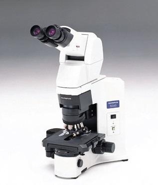 Figure 3 – BX45 microscope.
Sophisticated customer-based design
In the 2010s, Olympus improved the design and functionality of almost all of its microscopes, incorporating new features that focused on customer needs. The BX 53/43/46 microscopes were implemented with an LED light source that provides long life, high luminosity, and true-to-life color. Its fully ergonomic design provides improved user comfort and faster routine observation.
The IX inverted microscopes were updated with an insertable deck design to accommodate more diverse samples and a variety of applications in life science research. cellSens imaging software was developed with dual modes for high-end research image analysis as well as a simple and intuitive user interface for clinical users. Also introduced in this decade was the FV1200/1200-MPE laser scanning microscope, which featured higher sensitivity and faster imaging features.
The past decade has seen several notable achievements in the laboratory. More and more, researchers were using sCMOS (scientific CMOS) with extremely high quantum efficiency, and super-resolution microscopy, which pushed the limits of optical resolution. In the clinical environment, there was an increase in molecular- and gene-based diagnoses requiring a shorter turnaround time for each procedure.
Current technologies addressing today’s needs
As shown in Figure 4, a diverse range of samples can be imaged with next-generation Olympus microscopes. These include the FLUOVIEW FV3000 laser scanning confocal microscope, which was introduced in 2016 in response to the unique challenges of modern science. This microscope provides high-sensitivity multichannel imaging with macro to micro imaging capabilities, as well as improved productivity supported by high-speed imaging and a workflow-based intuitive user interface. The versatile frame of the FV3000 ranges from a simple, minimal configuration to customized advanced imaging.
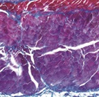
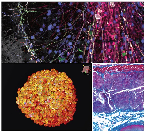 Figure 4 – Samples captured with FV3000 (top), IXplore Spin (bottom left), and DP74 (bottom right). Top: Brainbow AAV transfection of Purkinje cells, amplified with antibodies. Visible are Purkinje cell somata, dendrites, and axons, as well as some aspecific stainings of granule cells. Bottom left: cleared spheroid of HT-29 cells stained with DAPI (nuclear). Bottom right: Azan stained tissue.
In the past, Olympus provided advanced microscopes to researchers who focused on microscopic imaging. Today, researchers need to use a variety of techniques. Reflecting this trend toward multimodality, Olympus introduced the IXplore systems in 2017 (Figure 5). This series of solution-based preconfigured microscope systems enables researchers to choose the configuration best suited to their needs. There are six configurations dedicated to simple documentation, motorized multidimensional observation, precise live cell imaging, total internal reflection fluorescence (TIRF), spinning disk confocal, and super resolution.
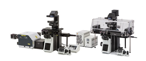 Figure 5 – IXplore solutions-based microscope system.
As technology advances, opening a world of deeper biological imaging, using super resolution to see beyond the optical limit has become a reality. SpinSR10 technology, a configuration of the IXplore microscope, provides fast confocal imaging with less phototoxicity and easy super resolution. The IXplore SpinSR10 microscope has modes for widefield fluorescence, confocal imaging, and super resolution, enabling researchers to delve into their samples faster, deeper, and more easily.
The BX53 microscope, launched in 2017 for use in clinical pathology, provides a level of color performance equal to that of a traditional halogen lamp, but with a newly developed, brighter LED. The microscope’s LED has an extended life of 50,000 hours, saving researchers money and down time. Further, the illumination control functionality of the BX53 synchronizes the brightness with the objective magnification, providing another way to achieve more comfortable and less time-consuming microscopic observation.
Conclusion
As a company founded on the goal of manufacturing world-class microscopes, Olympus continues to establish itself as a renowned precision technology leader, creating innovative opto-digital solutions in life science and beyond—including healthcare and consumer electronics products.
Today, Olympus’ “True to Life” spirit is a promise to deliver smart, creative solutions that change the world we live in and continue to revolutionize technology. Though it may be difficult to predict what the future holds for science and technology, as Olympus prepares to celebrate 100 years of contribution to these fields, it is certain that Olympus will be there.
Takeo Ogama is senior product and strategy planner, Olympus Corporation of the Americas, 48 Woerd Ave., Waltham, MA 02453, U.S.A.; tel.: 781-419-3619; e-mail: [email protected]; www.olympus-lifescience.com