Featured Article

Cell therapy and regenerative medicine have become firmly established as the newest branch of medical practice, and as a result preclinical research is heavily invested in the study of human primary cells and tissues. Cells isolated directly from human tissue rather than being grown in culture give researchers a much more biologically relevant model with which to study human diseases.1,2
Primary cells have historically been more difficult than cell lines to culture and propagate for more than a few days without loss of viability or cell type-specific function.3 In recent years, however, the virtual explosion of cell therapy research has fueled exciting advances in primary cell culture.4 The advent of 3-D culturing techniques, in particular, has been a boon to the primary cell industry, and these techniques are increasingly being used to support numerous biomedical research efforts.5-7
Apheresis, the practice of separating out and collecting various components of whole blood, is the primary source for the majority of primary cell starting material. However, variability is a significant issue with apheresis-derived material. Every donor is unique. Genetics, along with other key factors such as the donor’s medical background, can impact the quantity and functionality of various cell types in the donor’s circulation, which in turn will impact drug development research.8 The efficiency of traditional cell selection techniques both during and after apheresis determines how many target cells are in a given apheresis product, and therefore governs the functional potency of that product. Cell collection and isolation methods can also impact cell viability and cell health9 and lead to changes in cell subset populations and functionality.10
Researchers investigating the properties of a specific cell subset often need to request that donor material be collected in a certain way. For example, mobilization methods used to stimulate migration of hematopoietic stem cells into the peripheral blood of a donor affect the type of stem cells that are present in the apheresis product.11 Scientists considering various apheresis products for their research should ensure their supplier has the flexibility to source donor products collected via optimized mobilization and isolation protocols specific to a given project. Ideally, purity and functionality of the target cell type(s) can also be specified as required product quality control criteria.
Cell collection
While obtaining cells for an investigational project may seem straightforward, it is actually one of the most important decisions a researcher can make. Apheresis expertise, collection protocols, cell isolation, and handling methods all affect overall quality of the cellular material that supports biomedical research.
The most commonly used apheresis devices in the U.S. are the Spectra Optia (Terumo BCT, Lakewood, CO) and Fenwal Amicus (Fresenius Kabi, Lake Zurich, IL). While there are some hardware differences between the two, it is commonly agreed that cell collection efficiencies between them are equivalent. Use of the Spectra Optia is generally thought to result in lower red blood cell contamination.12
Assuming one has access to a strong apheresis donor network, collection techniques and predetermined quality control criteria will help ensure consistently high-quality material. Proper training and technique, particularly for specialized collection methods, are vital to the apheresis procedure.13
Beyond the procedure, programmed cell collection methods should be optimized for the blood component fraction or cell type that is the desired target of the collection. Collection of hematopoietic stem cells, for example, is guided by quality indicators such as CD34+ cell collection efficiency, which in turn is affected by the accuracy of optimal collection time predictions based on the stem cell mobilization method being used and the characteristics of the individual donor. The apheresis nurse must be prepared to accommodate changes in the programmed collection method to adjust endpoint collection time or volume and know how to manage any adverse events the donor may experience. Depending on the intended scientific goal, researchers may use the cell collection program to optimize either the volume or purity of the collected material.
Following collection of the apheresis product, further cell separation methods may be needed to isolate the specific cell type necessary to the research project. Positive and/or negative selection techniques are often carried out on a fluorescence-activated cell sorting (FACS) device using cell surface markers to guide the technician in optimizing the purity of the final product.14
Generally, researchers will want to set predetermined quality control criteria to determine whether the collected material is acceptable. Cell viability assays and functional assays may be carried out at the collection clinic to determine primary cell product quality, and that data is then passed on to the biomedical research lab.
Transferring apheresis products and primary cells to their intended destination is another critical quality determination point. No matter how high the initial quality of a cellular product, the entire point of the quality control procedure is lost if the integrity of the product cannot be safeguarded until it reaches the end user.
Cryopreservation of apheresis-derived primary cells is a common and logistically important way to protect the efficacy of these cells until they can be used for research. These methods must often be optimized to protect cell viability and functional integrity.15 A well-known example of this necessity is exemplified by the years-long debate over whether it was possible to cryopreserve mesenchymal stem cells (MSCs) and still retain their full function post-thaw.16,17 MSCs are highly valued in regenerative medicine for their capacity to differentiate and potential to treat graft versus host disease via their immunosuppressive properties. Without the option of safely cryopreserving the cells, however, it would be impractical to develop them for commercial manufacture and distribution. After extensive research into the problem, it was demonstrated that by modifying cryopreservation and thawing techniques, scientists could restore the full immunosuppressive capacity of these cells,18,19 and still take advantage of the valuable logistical flexibility conferred by cryopreservation.
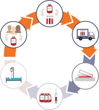 Figure 1 – Starting material impacts downstream processes: quality control systems protect downstream parameters such as data quality and product purity and potency. (Image credit: HemaCare.)
Summary
Apheresis material and primary cell providers should have quality systems in place to ensure that cell collection methods, instrumentation, training, and cell handling and processing procedures follow specified standard operating procedures that protect product quality, identity, and purity (Figure 1). Best practices in apheresis cell collection for biomedical and preclinical research depend on a number of key factors. Apheresis collection centers should maintain an extensive and diverse network of carefully screened, highly characterized donors who are amenable to repeat collections. Clinicians should be highly trained and qualified, and apheresis devices well-maintained according to standard operating procedures.
To ensure standards of quality, cell collection methods need to be guided by guaranteed quality indicators, such as optimal cell counts, high collection efficiency, and purity. Post-collection cell type-specific functional assays should be carried out both at the collection site and post-thaw for cryopreserved products. Establishing strict quality criteria and maintaining standard operating procedures during apheresis and cell collection will help protect primary cell product integrity and value.
References
- Hodgkinson, C.; Morrow, C.J. et al. Nat. Med. 2014, 20, 897–903.
- von Heideman, A.; Tholander, B. et al. Acta Oncologica 2014, 53(2), 242–50.
- Welser-Alves, J. ASCB Newsletter Nov 2015.
- Hussain, L. Gen. Eng. Biotech. News 2017, 37(13).
- Pageau, S.; Sazanova, O.V. et al. Biomaterials 2011, 32, 7169–80.
- Aljitawi, O.; Li, D. et al. Leuk. Lymphoma 2014, 55, 378–91.
- Drost, J.; Karthaus, W.R. et al. Nat. Protoc. 2016, 11, 347–58.
- Singh, N.; Perazzelli, J. et al. Sci. Translat. Med. 2016, 8(320), 320ra3.
- Grievink, H.W.; Luisman, T. et al. Biopreserv. Biobank. 2016, 14(5), 410–5.
- Worel, N.; Frank, N. et al. Transfusion 2017, 57(9), 2206–15.
- Baiamonte, E.; Barone, R. et al. Thalassemia Reports 2017, 7, 6392.
- Kim, J.; Joseph, R. et al. Transfusion and Apheresis Sci. 2016, 55(3), 368–70.
- Padmanabhan, A. Hematopoietic stem and progenitor cell collection by apheresis: techniques and tricks. In Hari, P. and Abutalib, S, Eds. Blood & Marrow Hematopoietic Cell Transplantation. Wiley Blackwell Publishers: Hoboken, NJ, 2017.
- Bruno, A.; Caravita, T. et al. Transfusion and Apheresis Sci. 2002, 26, 103–10.
- Woods, E.J.; Thirumala, S. et al. Cytotherapy 2016, 18, 697–711.
- Francois, M.; Copland, I.B. et al. Cytotherapy 2011, 14(2), 147–152.
- Moll, G.; Alm, J.J. et al. Stem Cells 2014, 32, 2430–42.
- Lauterboeck, L.; Hofmann, N. et al. Cryobiology Dec 2015, 71(3), 384–90.
- Pollock, K.; Samsonraj, R.M,.et al. Stem Cells and Devel. 2017, 26(11), 828–42.
Dominic Clarke, Ph.D., is global head of Cell Therapy, and Marie Aragon, RN, BSN, is Donor Center director, HemaCare Corp., 15350 Sherman Way, Ste. 423, Van Nuys, CA 91406, U.S.A.; tel.: 877-310-0717; e-mail: [email protected]; www.hemacare.com