Featured Article

Preclinical imaging (PCI) is vitally important for studying the biological processes behind disease states at the organ, tissue, cell, and molecular level. Revealing how the body responds to physiological or environmental change drives the search for therapeutic agents to combat disease. The efficacy and safety of new treatments is also evaluated by PCI to inform researchers of drug distribution patterns in tissues. Tomography—an imaging technique used across a wide variety of fields including radiology, nuclear medicine, geophysics, and materials science—provides three-dimensional information about a subject based on its sections or projections. Common examples include X-ray, computed tomography (CT), positron emission tomography (PET), and single-photon emission computed tomography (SPECT).
CT scanning provides information on the anatomy of the subject, while PET offers functional imaging that shows the spatial distribution of biomolecular activity in the body. PET was developed as a technology for both clinical diagnostic and preclinical purposes in the 1950s, the scope of which was expanded by the development of radiopharmaceuticals—a group of pharmaceutical drugs that emit radiation and commonly includes radiotracers. PET in preclinical studies enables users to conduct repeat experiments on the same animal subjects, providing strong, statistically valuable data, and thereby reducing the number of animals required for a study. For this reason, it has become increasingly important to use noninvasive in vivo imaging techniques to optimize the use of each animal used.
Multi-modal imaging
Multi-modal tomographs, such as PET/CT, correlate the functional imaging obtained using PET with the anatomic imaging obtained with CT scanning. PET can also be combined with other technologies, such as magnetic resonance imaging (MRI), to bring functional imaging together with soft tissue morphological imaging. Preclinical imaging applications are increasingly looking toward PET/MR for its superior soft tissue contrast, imaging without the CT’s ionizing radiation risk, and multiparametric data. Multi-modal imaging technologies such as PET/CT, SPECT/CT, PET/MR, and PET/SPECT/CT are useful in a variety of research applications, including oncology and cardiology.
Monitoring tumor development
Many cancers are associated with a higher metabolic turnover than normal cells. Glucose uptake can be quantified by using PET and an injected radiolabeled glucose analogue tracer such as fluorine-18 (18F)-fludeoxyglucose (18F-FDG). This method can also be used to detect tumor burdens and molecular biomarkers to contribute to cancer detection and treatment response assessment. PET/CT, and more recently PET/MR, are used to determine the accumulation regions of 18F-FDG to obtain a semiquantitative standardized uptake value (SUV) to assist in the diagnosis of tumor malignancy.
Combination cancer therapies are often desired for their ability to address multiple molecular targets as well as a reduced chance of drug resistance. Preclinical PET/CT imaging was used to monitor 18F-FDG tumor uptake for different treatment combinations: radiotherapy (Rad) alone, Rad + Temozolamide (Tmz), Rad + Mifepristone (Mife), and Rad + Mife + Tmz. Rad+ Tmz is the typical treatment regime for glioblastoma, but the study found that using Mife as a priming agent suppressed tumor growth more than the other treatment combinations1 (Figure 1). The mechanism of this chemo-radio-sensitizing effect of Mife is yet to be fully characterized, but studies such as this help researchers make important steps toward improving available cancer treatments.
Preclinical laboratories are increasingly aware of the benefits of PET/MR for oncology research. As a result of MRI’s unique ability to image soft tissue, users can visualize the true tumor margin and evaluate tracer distribution within individual tumors to generate a desired volume of interest (VOI) and calculate SUVs more accurately. Tumor margin detection is a unique and significant enhancement to preclinical cancer PET studies.
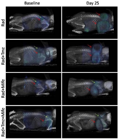 Figure 1 – PET/CT images showing 18F-FDG tumor uptake, in four treatment combinations, at the beginning of treatment and 25 days later. Red arrows indicate tumor location at baseline and day 25; green arrows show sites of typical 18F-FDG uptake in brown adipose tissue (BAT). (Reproduced from Ref. 1 in accordance with the Creative Commons License, https://creativecommons.org/licenses/by/2.0/).
Cardiology
Preclinical researchers use multi-modal PET imaging to gain a deeper understanding of cardiovascular metabolism, inflammation, and perfusion. High spatial and temporal resolution cardiac imaging is crucial for resolving features of cardiac disease, including plaques and ischemic lesions, in small animals. The primary assessment parameter for cardiac preclinical imaging is myocardial metabolic viability and perfusion, but others such as inflammation, innervation, apoptosis, and neovascularization are also attainable through PET.2
MRI, PET, and microCT imaging are well-established in the study, diagnosis, and treatment of heart disease. Myocardial PET is used to separate cardiac disease stages from normal physiological stages, which is important in the evaluation of therapeutic strategies, and the determination of imaging biomarkers of coronary artery disease (CAD). Resolution is an important factor in cardiac imaging, because blood concentration is often obtained from the left ventricle. The higher the resolution, the lower the partial volume effect and, therefore, the lower the chance of contamination from the myocardium into the blood concentration derived from imaging. PET scanners that maintain resolution regardless of the animal’s position in the field of view (FOV), such as PET systems from Bruker (Billerica, MA), open up the possibility of multi-animal imaging, enabling researchers to obtain results with low or ultralow tracer activity, at high diagnostic accuracy, in the shortest possible time frame.
Sophisticated preclinical PET scanners can achieve submillimeter resolution and allow accurate detection of small lesions as well as better characterization of the right myocardial physiology. These scanners are also able to image smaller animals, such as mice, in models that were previously only performed in rats, adding the valuable use of genetically modified rodent strains. The CT capabilities of state-of-the-art PET/CT scanners provide complementary information to PET imaging, such as calcification area and fine shape of vessel and cardiac structure in murine models. Although PET/CT is considered the “gold standard” for cardiac imaging, PET/MR is gaining more ground in this area of preclinical research. MRI information can be used for retrospective cardiac/respiration gating (IntraGate, Bruker BioSpin), without the use of electrocardiogram (ECG) electrodes, and PET/MR fingerprinting can provide information on tissue metabolic state, structural integrity, perfusion, global/local function, and molecular pathways in the same subject.
PET/MR is particularly beneficial for plaque imaging and the molecular characterization of inflammation, making the technology useful for characterizing disease states associated with these factors. PET/MR is also suitable for the in vivo monitoring of new drugs being developed to target inflammatory diseases. PET is one of the few technologies capable of delineating cardiac autonomic denervation, the degree of which has been shown to identify those at risk of cardiovascular events.3
Technological developments
In order to provide the highest-quality imaging results, PET systems must meet a range of performance criteria. The material in the detector stopping the gamma rays (scintillator), as well as detector design, influence the resolution and sensitivity of images. The size of scintillator crystals impacts PET resolution, and dense, thick crystals are often the material of choice in PET detectors due to their ability to stop as many gamma rays as possible.
Ring diameter and depth of interaction (DOI) correction must also be optimized for high-resolution imaging. PET sensitivity is also determined by crystal technology, which has undergone rapid development in recent years. In contrast to the traditionally used pixelated crystals, continuous crystals have shown to better measure light distribution and vastly improve resolution and sensitivity.
Novel light detection technology has been combined with continuous crystal scintillators in new, advanced PET systems for unparalleled imaging capabilities. In such systems, continuous crystals are coupled with silicon photomultiplier (SiPM) photosensors for accurate determination of all three spatial coordinates of the gamma–photo interaction within the detector crystal, resulting in submillimeter spatial resolution, regardless of the positron. This is known as full field accuracy (FFA) and results in more reproducible data, independent of variable sample positioning. It also provides more reliable imaging of large samples, or multiple animals across the FOV, increasing accuracy and throughput in preclinical imaging. SiPM technology is also becoming increasingly popular for its compatibility with strong magnetic fields employed by MR.
Future directions
As a highly sensitive, noninvasive technique, PET can help improve the understanding of the underlying causes of disease, expanding methods of detection and treatment. Preclinical PET studies facilitate the development of imaging biomarkers, with the goal to translate these to the clinic to identify patients at risk or in the early stages of disease. Multi-modal systems make it simple for researchers to combine the benefits of PET imaging with CT, SPECT, and MRI technology for optimal imaging results.
References
- Llaguno-Munive, M.; Medina, L.A. et al. Mifepristone improves chemo-radiation response in glioblastoma xenografts. Cancer Cell International 2013, 13, 29; https://doi.org/10.1186/1475-2867-13-29.
- Nekolla, S.G.; Rischpler, C. et al. Cardiovascular preclinical imaging. Q. J. Nucl. Med. Mol. Imaging 2017, 61, 48–59; doi: 10.23736/S1824-4785.16.02960-5.
- Schwaiger, M.; Kunze, K. et al. PET/MR: Yet another Tesla? J. Nuclear Cardiol. 2017, 24, 1019–31; doi: 10.1007/s12350-016-0665-2.
Sonica Van Wyk is market product manager, Nuclear Molecular Imaging, Bruker BioSpin, Preclinical Imaging Division, 15 Fortune Dr., Billerica, MA 01821, U.S.A.; tel.: 647-326-2559; e-mail: [email protected]; www.bruker.com