Featured Article

Modern molecular biology offers a variety of ways to investigate the behavior and properties of cancer cells. Application of cell culture in cancer research allows scientists to identify novel drug targets and preliminary testing of new drugs. Particularly, with high-throughput assays, scientists can screen large substance libraries for new active substances that target specific cancer cell receptors. Drugs based on newly developed modes of action are suitable for a well-defined group of patients, transforming cancer therapy into personalized medicine.
One concept that is agreed upon in the scientific community is that no two types of cancer are the same. Different cells require conditions that are specific to their needs. Herein lies one of the largest flaws of in vitro cancer research. While it is fully understood that tumor cells rely heavily on their microenvironment for growth, most researchers still grow their cells, regardless of type, in an undefined growth medium and within incubators that are under normoxic (18–21% O2) conditions. This way of growing cells is far from what would be expected under any in vivo circumstance.
This article delves into these microenvironments further and suggests, based on the authors’ current understanding, that researchers should be cognizant of controlling for these factors. It will first investigate how most growth media used by cancer researchers today is not physiologically relevant and what options exist to provide researchers with more appropriate growing conditions. The latter half of the article focuses on the external growth factors and why more researchers should consider controlling oxygen levels, which can significantly impact cellular behavior. This article should enhance the researcher’s understanding of the optimal growth conditions to which cancer cells should be subjected in vitro to better resemble the in vivo experience they are being studied for.
In vitro immune cells and growth media
Modern cell cultures, like minimum essential medium (MEM), were developed over 60 years ago, yet most in vitro cell cultures continue to utilize these basic formulations combined with bovine or human serum for experimentation. Cell culture has become even more important over the last decade as cellular and gene therapies, in which the patient’s own cells are isolated and grown in vitro, have proven to be an important advancement for cancer treatment in the clinic. These therapies continue to use cell culture media that have become standard in the industry with little thought as to how the medium may be affecting the cells while in culture. When comparing DMEM to physiological ranges in the human body, DMEM contains four times the normal amount of glucose.1 This increased amount of glucose has been found to cause altered gene expression, protein production and function, as well as increased osmotic and oxidative stress that can be detrimental to cells. Another popular medium, RPMI 1640, contains very low levels of calcium, magnesium, and sulfate, but has three times the normal level of phosphate. Some popular basal medias have proprietary formulas that are unable to be compared to human physiological levels; therefore, the consumer has no inclination of what the cells may be exposed to in culture. The same can be said for human serum (HS) and fetal bovine serum (FBS), which contain a plethora of unknown contaminants that cause high variability and can be seen as a potential source of harmful infectious agents. For these reasons, regulatory bodies such as the Food and Drug Administration (FDA) and European Medicines Agency (EMA) are discouraging the use of these animal products. Chemically defined media not only remove the need for human or animal serum, but allow the researcher to control the components the cells are exposed to and may be able to increase cell growth, control phenotype, or tailor the cell behavior to fit their needs (Figure 1).
 Figure 1 – InVitria’s OptiPEAK T Lymphocyte Medium is a chemically defined blood free expansion medium specifically optimized for human T lymphocytes.
An article by Ed Yong2 argues that off-the-shelf media may be skewing results due to the unnatural levels of sugars, salts, vitamins, and amino acids. The article illustrates the benefits of using a medium that mimics the constitution of human blood so that the cells may behave more like they would naturally in the human body. However, a medium that is consistent with that of human blood may not be representative of that in tumor environments, which are known to be hypoxic and have different nutrients available than that of healthy plasma. N-acetylcysteine (NAC) is one example of a component that could prove to be beneficial in certain cell culture systems. Research shows that the addition of NAC in peripheral blood T cells increase cell expansion and interleukin-2 (IL-2) production.3 As immunotherapy research progresses to target solid tumors, researchers should also account for the differences between tumor in circulation versus in mass.
Different cell types also have significantly different growth requirements. Chemically defined media can provide control over the cell type and end intention that the researcher may be targeting. Furthermore, cells from a healthy patient can have a very different metabolic profile than that of a diseased patient. Off-the-shelf media may be developed using healthy models that would not fully capture the needs of diseased cells. Chemically defined media can be formulated to meet the needs of the specific cell type and disease indication without the variability and supply chain shortcomings that come with using proprietary growth medium and animal products such as human or fetal bovine serum.
In vitro cancer research and the external environment
One of the greatest developments in keeping, growing, and culturing cells has been the advancement of laboratory incubators to maintain cell cultures over time. CO2 incubators have been commercially available for over 60 years and provide control over crucial factors such as temperature, CO2 (for maintaining proper pH levels), and humidity, all of which affect cell growth. The purpose of these chambers is to essentially mimic the in vivo experience that cells face, allowing them to flourish in a “natural” environment. This continues to be the standard for cancer researchers to grow their cells. To fully mimic the in vivo microenvironment, many researchers are beginning to stress the importance of controlling, arguably the most important external physiological growth factor, oxygen.
The lack of research taking oxygen into account is quickly becoming a contested issue across the entire cellular research community. Depending upon the physiological or pathological context, oxygen levels can vary widely within the body, with most tissues falling between 5 and 12% oxygen. CO2 incubator air exposure is approximately 18% oxygen and is contradictory with physiological in vivo conditions.
Within cancer, specifically within tumor formation, highly hypoxic environments (>2% O2) are the norm, and severely augment cell function. Hypoxia is a common characteristic of locally advanced solid tumors that is accompanied by a diminished therapeutic response. For example, cells specifically designed to fight cancer, like CAR-T cells, under hypoxia are often diminished in expansion, differentiation, and cytokine production, which contributes to the inability of CAR-T cells to eradicate solid tumors.4 Further, hypoxic environments also lead to greater malignant progression, increasing the probability of recurrence, distant metastasis, and locoregional spread of tumors. Emerging evidence indicates that the effect of hypoxia on malignant progression is mediated by a series of hypoxia-induced proteomic and genomic changes activating angiogenesis, anaerobic metabolism, and other processes that enable tumor cells to survive or escape their oxygen deficient environment.5
The transcription factor hypoxia-inducible factor-1 (HIF-1) is the major regulator of tumor cell adaptation to hypoxic stress. Tumor cells with proteomic and genomic changes favoring survival under hypoxic conditions will proliferate, thereby further aggravating the hypoxia. The selection and expansion of new (and more aggressive) clones, which eventually become the dominant tumor cell type, leads to the formation of a vicious circle of hypoxia and malignant progression. Cancer researchers growing cells in CO2 incubators at 18% will completely miss these conditions, making it difficult to assess any in vivo circumstance accurately. There are numerous pathways greatly augmented by HIF (including metabolism, angiogenesis, tumor progression and resistance, cell death, and anemia) and, if not controlled for, can alter the outcome of any cellular study. This is even more pertinent to cancer research as tumors often reside in ultra-low oxygen conditions.6
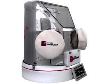 Figure 2 – Oxford Optronix’s HypoxyLab is a standalone benchtop hypoxia incubator and workstation that controls all standard culturing conditions (CO2, temperature, and humidity) while regulating O2 levels.
For most researchers, the simplest way to mitigate this issue is through the use of a hypoxia (tri-gas) incubator or workstation that can provide an in vivo-like O2 atmosphere for cells. This completely removes much of the doubt as to whether oxygen played any role in a study and inevitably increases the accuracy of all results. There are hypoxia chambers available in all sizes to meet the needs of any research group. From standard benchtop units (Figure 2) to adding hypoxic elements into standard CO2 incubators (Figure 3), cancer researchers have more options than ever to work under physiologically relevant conditions.
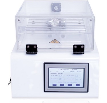 Figure 3 – Baker Ruskinn’s PhO2x Box regulates O2 and CO2 while using the set temperature and humidity from a standard culturing incubator.
Conclusion
Cellular research is the foundation of biomedical innovation for all types of cancer. The lack of research using relevant media and oxygen levels in cellular cancer studies is quickly becoming a highly contested issue across this large research community. Basic research using chemically defined media and oxygen regulating chambers can help researchers begin to study the human immune system with more direct correlations between cells grown inside and outside the human body. These fundamental changes can be instrumental in understanding the conditions in which our immune systems work and speed up the development of novel therapies for targeting cancer.
References
- McKee, T.J. and Komarova, S.V. Is it time to reinvent basic cell culture medium? Am. J. Physiol.-Cell Physiol. Feb 2017.
- Yong, E. Scientists have been studying cancers in a very strange way for decades; https://www.theatlantic.com/science/archive/2019/01/cancer-culture-media-plasmax/579283/
- Eylar, E.; Rivera-Quinones, C. et al. N-acetylcysteine enhances T cell functions and T cell growth in culture. Int. Immunol. Jan 1993.
- Berahovich, R.; Liu, X. et al. Hypoxia selectively impairs CAR-T cells in vitro. Cancers Apr 2019.
- Keeley, T.P. and Mann, G.E. Defining physiological normoxia for improved translation of cell physiology to animal models and humans. Physiol. Rev. Oct 2018.
- Vaupel, P. The role of hypoxia-induced factors in tumor progression. The Oncologist Nov 2004.
Justin R. Croft, MSc, is application specialist—Cell Biology, Scintica Instrumentation, 562 Waterloo St., London, ON, N6B 2P9, Canada; tel.: 519-914-5495; e-mail: [email protected]; www.scintica.com. Annie Cunningham is cell culture scientist, InVitria, 12635 East Montview Blvd., Aurora, CO, 80045, U.S.A.; tel.: 1-800-916-8311; e-mail: [email protected]; www.invitria.com.