Featured Article
The following is a report on an important technology featured at the 67th ASMS Conference on Mass Spectrometry and Allied Topics, held in June in Atlanta, GA.
IonDx (Monterey, CA) presented a poster introducing a benchtop ion mobility spectrometer (IMS) designed for separation and characterization of larger biomolecules and polymers. Since the IMS separation unit is based on a simple metal collection ring rather than a mass spectrometer, the instrument’s price point is much lower. Plus, it is optimized to separate polymers in their native state.
This is a neat trick, since the polymers are ionized in an electrospray ion source (ESI), which usually produces multiply charged ions. The molecular ion often rearranges itself to distribute the charges all over the ion, which can lead to rearrangement of the structure. The secret sauce is to put a poly-ionic neutralization stage immediately downstream of the ESI source. In this case, it is a radioactive polonium source that provides a cloud of low-energy bipolar air ions that reduce the charges on electrospray droplets, allowing singly charged ions to float away having never passed through a high charge state. The mono-charged ions are resolved with the ion optics shown in Figures 1 and 2.

Figure 1 – The sample is pumped through an electrospray capillary and the resulting gas-phase ions enter the spectrometer through the center bore of a bundle of tubes that guide the flow toward a center rod. In conventional ESI sources, the electrospray droplets dry and fission, then pass to a mass spectrometer. In the case of the IonDx instrument, a collection of multiply charged ions pass through the grounded screen forming the counterelectrode and into the drift region created by voltage applied to the centered rod (right, Figure 1). The conducting ion collector ring is placed downstream of the ground grid.

Figure 2 – Comparison of normal electrospray ionization (upper figure), which produces multiply charged ions upon desolvation, giving rise to different proteoforms. The lower figure shows singly charged ions resulting from placing a 210Po source in the plume of the ESI source. These do not have energy to rearrange. Preserving the native conformation is useful in characterizing large biomolecules such as antibodies, antibody drug conjugates, and possibly prions, fibrils, and plaques. After passing through the counterelectrode grid, the analyte ions from the ESI immediately experience the strong repulsive electrical field created by the high voltage from the center electrode. If the ion has a small collisional cross-section (CCS), such as the solvent (blue line), and the same polarity as the rod, it quickly moves to the side wall and is lost. If the ion is too large (red path), it over-flies the detector ring and is passed down the tube and lost. Only ions that have the selected CCS hit the detector ring, thus producing an electrical current, which is amplified and recorded.
However, as shown in the top section of Figure 2, the ions from ESI usually have multiple charges. The charges can be reduced when a radioactive 210Po source is inserted immediately downstream of the ESI Taylor cone. Polonium decay produces an extremely high concentration of negatively and positively charged air ions that are attracted to and neutralize nearly all of the positive charges on the positively charged electrospray droplets. In fact, this charge neutralization mechanism is reproducible and produces singly charged droplets in the electrospray source. These ions are dispersed to the detector ring from which the ion current is measured. The charge-reduced drops from the ESI source do not experience the highly charged state illustrated in the top of Figure 2. This preserves the native state configuration of the sample, which is measured by the IonDx spectrometer.
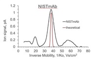
The voltage applied to the center rod (Figure 1, right) can be constant or ramped with time. The latter permits scanning the mobility of the ions. The normal voltage range is scanned from 0 to 3 kV in 90 seconds. A typical output is presented in Figure 3.
The peak is wider than predicted for a monostructure of the NISTmAB standard, which may be related to the microheterogeneity of the NIST reference standard.
Figure 3 – IMS of NISTmAB standard using the DX instrument. MW ~150,000 Da. The increase in bandwidth is due to instrumental and microheterogeneity of the sample
The authors concluded their poster with:
“The Dx ion mobility spectrometer is capable of measuring the mobility constant (Ko) of singly charged ions that have not experienced high charge states and may be more representative of solution structure.
The average conformation in terms of CCS [collisional cross-section] can be extracted from the position of the peak and the width of the ion mobility peak represents the extent of polydisperity.”
Antibody aggregates could be dumbbell-shaped.
Future applications:
“We will present thermograms for several biotherapeutic antibodies including NISTmAb and use these plots to calculate melting point temperatures. Only 0.5 µg of sample is needed, thus allowing thermal stability tests to be run at all stages of product development, including early stages when bacterial expression vectors are used before manufacturers switch to mammalian expression systems. We will discuss extending this technology to process monitoring, including automation based on 96-well sample plates.
This instrument is being used by a major commercial clinical diagnostics firm to measure the conformation of some diagnostically significant protein biomarkers. We expect more assays will be developed as the number of spectrometers increases.”1
Reference
- Benner, W.H. and Aguilar, B. A New Bench-top Dispersive Ion Mobility Spectrometer for Characterizing Biotherapeutic Drugs. IonDx, Monterey, CA. Poster ThP 275, ASMS 2019.
Robert L. Stevenson, Ph.D., is Editor Emeritus, American Laboratory/Labcompare; e-mail: [email protected]