Over the last several years, cryo-electron microscopy (cryo-EM) has revolutionized structural biology by enabling scientists to obtain high-resolution models of molecules that could not be uncovered using other approaches. Cryo-EM was named Nature Method of the Year 2015, and three luminaries in the cryo-EM field won the Nobel Prize in Chemistry 2017 for their foundational work on this technique.
Today, cryo-EM has become instrumental in drug discovery in areas ranging from cancer to neurological disorders to infectious diseases. In 2020 alone, discoveries using cryo-EM to study the novel coronavirus made their way into more than 80 publications.
While cryo-EM has generated remarkable breakthroughs, until recently the technology was both expensive and complex to use, requiring organizations to build special rooms to house, and hire dedicated experts to operate and maintain these instruments. New cryo-EM users faced lengthy learning curves, typically requiring at least a month to obtain their first structure as they navigated complex workflows involving thousands of small steps. What’s more, many institutions couldn’t afford these large instruments or the cost of new facilities needed to house them, limiting access only to well-funded pharmaceutical companies and academic institutions.
Extending Cryo-EM to More Users
Thermo Fisher Scientific has been working to break down these barriers to adoption by designing a transmission electron microscope (TEM) that democratizes cryo-EM. The Thermo Scientific Tundra Cryo-TEM delivers on this promise as a cost-effective instrument that extends cryo-EM to users of all experience levels.
Tundra Cryo-TEM incorporates artificial intelligence and automation to help non-experts navigate complex workflows. Results are displayed in a “traffic light” style with a green light indicating that users can proceed and a red light notifying them when something is wrong, while offering guided instructions to help them quickly fix the problem. Tundra also provides an integrated loader that makes it easy to load samples into the microscope. Users can exchange sample carriers in about two minutes, enabling them to rapidly optimize sample conditions as they check their results.
Tundra delivers this unprecedented ease-of-use without trade-offs in image quality or performance. The instrument generates resolutions as high as 3.5 angstrom, with throughput of 24 to 72 hours per dataset. Novice users can typically begin obtaining quality results on day one of operation, instead of having to go through extensive training.
Offered at a significantly lower price point than conventional cryo-TEMs, Tundra is making it possible for a much wider swath of academic institutions and pharmaceutical companies to obtain structural insights at biologically relevant resolutions. And it delivers a compact footprint that
fits standard-sized laboratory rooms, reducing the need for expensive renovations.
Taking Disease Research to the Next Level
To advance our knowledge of the diseases – both old and new – that impact the human race, it’s critical to obtain a detailed understanding of the key proteins that play a role in cellular function. Yet many of the tools scientists used in the past to research protein structures could only study proteins indirectly or in isolation, limiting their ability to observe proteins in different states or with glycan modifications, or identify the complex ways in which individual proteins interact with one another.
Cryo-EM solves these problems by enabling researchers to see biological mechanisms in their near-native states, including the different shapes and forms proteins take on as they interact with one another. It’s these details that lead to life-saving treatments, such as better drugs with fewer side effects.
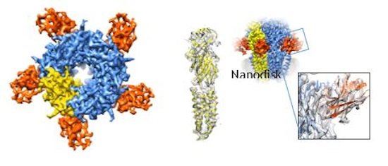
Figure 1: Using Tundra Cryo-TEM, researchers obtained a 4.3 angstrom resolution structure of the GABAA small membrane protein with bound megabodies in its near-native state. (Image courtesy of Radu Aricescu, MRC LMB, Cambridge, as well as Dimple Karia and Abhay Kotecha, Thermo Fisher Scientific.
For example, the human gamma-aminobutyric acid type A receptor (GABAA) is a small membrane protein and ligand-gated chloride-ion channel that mediates inhibitory neurotransmission. GABAA receptors are important therapeutic targets as their various conformations affect a variety of important signaling pathways. Yet scientists had been unable to crystalize this flexible and dynamic receptor. Using cryo-EM, they have been successful in resolving the structures of the physiological forms of this receptor, helping to understand its mechanism of action.
Using Tundra, researchers obtained a 4.3 angstrom resolution of the GABAA structure with data collected in just three days. At this resolution, every turn and strand of alpha helix and beta sheet secondary structure is resolved, and important functional details of protein-protein interactions (for example, in antibody epitope mapping) are readily visible.
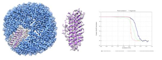
Figure 2: Using Tundra Cryo-TEM, researchers obtained a 3-angstrom resolution structure of the rigid benchmark protein apoferritin with data collected over just 1.5 days, enabling scientists to clearly visualize bulky amino acid side chains and build atomic models.
Similarly, apoferritin is an important protein commonly present in the intestinal mucosa and the liver. With Tundra, researchers obtained a 3-angstrom resolution structure of apoferritin in just one-and-a-half days, enabling scientists to clearly visualize the protein backbone and major amino acid side chains needed to build de novo models. Data at these resolutions can help scientists to understand how proteins function, how to modify genes, and how to design drugs accordingly.
Accelerating the Pace of Cryo-EM Discoveries
By reducing the cost and complexity of cryo-EM, the goal is to put this innovative technology into the hands of more scientists. Already, cryo-EM is playing a lead role in many of the discoveries transforming our understanding of human diseases. And with easier and wider access to this important technique, cryo-EM research is poised to expand, accelerating the pace of critical breakthroughs—and ultimately the development of highly effective drugs and vaccines.
References:
- Phulera, S., Zhu, H., Yu, J. et al. Cryo-EM structure of the benzodiazepine-sensitive α1β1γ2S tri-heteromeric GABAA receptor in complex with GABA. eLife 7, e39383 (2018). https://doi.org/10.7554/eLife.39383
- Liu, S., Xu, L., Guan, F. et al. Cryo-EM structure of the human α5β3 GABAA receptor. Cell Res 28, 958–961 (2018). https://doi.org/10.1038/s41422-018-0077-8
- Laverty, D., Desai, R., Uchański, T. et al. Cryo-EM structure of the human α1β3γ2 GABAA receptor in a lipid bilayer. Nature 565, 516–520 (2019). https://doi.org/10.1038/s41586-018-0833-4
- Masiulis, S., Desai, R., Uchański, T. et al. GABAA receptor signaling mechanisms revealed by structural pharmacology. Nature 565, 454–459 (2019). https://doi.org/10.1038/s41586-018-0832-5
- Nakane, T., Kotecha, A., Sente, A. et al. Single-particle cryo-EM at atomic resolution. Nature 587, 152–156 (2020). https://doi.org/10.1038/s41586-020-2829-0