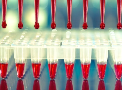
The preferred source of DNA in human genetics research is blood, or cell lines derived from blood, as these sources yield large quantities of high-quality DNA. However, DNA extraction from saliva can yield high-quality DNA with little to no degradation/fragmentation that is suitable for a variety of DNA assays.
In fact, saliva and buccal samples have become increasingly valuable sources of genetic material for clinical applications due to ease of access (non-invasive sample collection), as well as convenient storage and transport procedures. Multiple reports indicate that saliva samples provide better quality DNA than buccal samples.1
But, extracting DNA in the laboratory isn’t always as straightforward as lyse, precipitate, wash, suspend, especially given today’s increasingly advanced DNA analysis methods like multiplex, real-time PCR and next-generation sequencing.
1. Optimize critical parameters
The amount and quality of DNA obtained from human samples can vary widely depending on the state and type of starting material. In general, best laboratory practices involve minimizing the amount of time between the removal of the bodily fluid or tissue from the human body (or cell culture dish) and storage at a very low temperature (−80°C or liquid nitrogen storage highly recommended).1
For Formalin-Fixed Paraffin-Embedded (FFPE) tissue samples, it is critical to optimize the fixation conditions of each tissue type to not over-fix the tissue in the formalin. This will make downstream DNA isolation much more difficult and result in lower yield.1
When using a kit for DNA extraction, is especially important to note the expiration date of the kit since many of the wash buffers contain ethanol, which can evaporate and react with water, and result in less-effective washing. Before using DNA in downstream assays, it is highly recommended that the DNA concentration be determined using a fluorometric assay, as this is the most accurate method.1
2. Avoid contamination with specialized lab areas
Obtaining a clean, successful PCR requires samples free of exogenous DNA. But contaminating DNA can be lurking around every corner—from previously amplified products hanging out in the lab to your own DNA.
To avoid common types of contamination, designate and use distinct areas for sample preparation, PCR setup, and post-PCR analysis. To eliminate contamination from old amplicons, set up the stations on separate benchtops, one for pre-PCR (for PCR reaction setup only) and the other for post-PCR (purifying PCR-amplified DNA, measuring DNA concentration, running agarose gels, and analyzing PCR products). Restrict equipment to these areas—keep the PCR machine and electrophoresis apparatus in the post-PCR area. This is the golden rule of PCR: do not bring any reagents, equipment, or pipettes used in a post-PCR area back to the pre-PCR area. This even goes for lab notebooks and pens. 2
Additionally, prepare and store reagents for PCR separately and use them solely for their designated purpose. Aliquot reagents in small portions and store them in either location based on their use in pre-PCR or post-PCR applications.2
Use separate sets of pipettes and pipette tips, lab coats, glove boxes, and waste baskets for the pre-PCR and post-PCR areas. If you're doing NGS library prep, use a surface decontaminant for nucleic acids to wipe down benchtops and pipettes before beginning. Moreover, be sure to use pipettes and pipette tips with aerosol filters dedicated for DNA sample and reaction mixture preparation.2
Whenever possible, keep the number of PCR cycles to a minimum as highly sensitive assays are more prone to the effects of contamination. Lastly, be sure to detect PCR contamination before it has the change to affect downstream applications. Be sure to perform a negative control reaction that omits template DNA, substituting it with ultrapure water to ensure no contamination has occurred.2
3. Consider automation for purification
Automation is another way to significantly reduce the risk of cross-contamination between samples, in addition to enhancing efficiency and robustness of extractions. Nowadays, many researchers use magnetic bead–based kits for nucleic acid extraction due to their ease of use and reliability of results. The protocols for magnetic bead–based sample preparation kits include steps in which reagents must be dispensed into 24- or 96-well plates. While researchers can manually pipet samples onto the plates, it is tedious, time-consuming, prone to error, and inefficiency. On the other hand, automated systems reduce inconsistencies in sample yield, preparing uniform quantities for PCR applications and sequencing analyses.
In a recent technical note, Thermo Fisher Scientific evaluated the efficiency and reproducibility of manually dispensing magnetic beads and extraction reagents compared with dispensing them with the Thermo Scientific Multidrop Combi Reagent Dispenser. The timing results showed that dispensing with the Multidrop Combi dispenser was more efficient than manual dispensing, reducing the time by 62 to 73% when used for dispensing magnetic beads and reagents. Additionally, the A260/A280 ratios for the DNA extractions were between 1.79 and 1.85 for both dispensing methods, indicating that the samples were clean and free from carry-over contaminants.
Automation is also an ideal solution for researchers who work with rare or difficult-to-obtain samples—such as forensic DNA, patient biopsy tissue or even historical samples—as it conserves starting sample material. It reduces the risk of human errors resulting in loss of sample or the need to redo the sample prep process.
Of course, automation is not for every laboratory. When considering the move from manual to automated sample prep, take into account the number of samples processed daily and available laboratory space. In contrast to low throughput manual kits, automated DNA and RNA purification systems can process 12 to 96 samples in less than 30 minutes or 1 hour, respectively, and allow various handling volumes ranging from 1 to 1000 µL.
References
1. Miskimen, K. L. S., & Miron, P. L. (2023). Isolation of genomic DNA from mammalian cells and fixed tissue. Current Protocols, 3, e818. doi: 10.1002/cpz1.818
2. "7 tips for avoiding DNA contamination in your PCR." Takara Bio. (July 30, 2018). https://www.takarabio.com/about/bioview-blog/tips-and-troubleshooting/avoid-dna-contamination-in-pcr