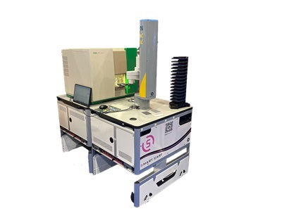
Custom flow cytometry workcell featuring the ZE5 Cell Analyzer with the KX-2 Laboratory Robot and S-RUN Laboratory Automation software.
by Richard J. Cuthbert, PhD, Product Manager, Bio-Rad Laboratories
High-throughput screening (HTS), using automated equipment to rapidly test thousands to millions of samples for biological activity,1 is central to drug discovery. Flow cytometry is now a key component of the HTS toolbox, being instrumental in antibody and phenotypic screening all the way up to lead optimization. High-throughput flow cytometry offers an unparalleled combination of speed, sensitivity, and multiparameter analysis for use in both pharma and biopharma, with applications spanning every stage of the drug discovery and development process.
Generating a highly specific biologic, such as a novel antibody-based drug, often involves high-throughput library screening of hundreds of candidates to find those that bind strongly and specifically to target antigens with the potential to become commercial products. For small molecule drug discovery, phenotypic screening employing a target-agnostic approach is used to evaluate molecules based on their ability to promote a desired phenotype associated with a therapeutic effect. These contrasting screening approaches, as well as lead optimization where the favorable characteristics of promising molecules or biologics are refined and enhanced, have much to gain from the latest advances in high-throughput flow cytometry.
Ongoing research and development continue to drive the considerable benefits of this technology forward with impressive progress being made in automation and robotics to enhance throughput capabilities. In this era of great innovation, researchers need to think carefully about which flow cytometry platform will best suit their screening requirements.
Increasing speed, reducing sample volume
Traditionally, flow cytometers relied on drawing one sample at a time through a section of capillary tubing before passing each particle through an illumination source. This approach required the use of large sample volumes and cleaning between samples. Developing a technique that allowed multiple samples to be drawn sequentially into the sample line (each separated by air or water to distinguish between samples) was a major breakthrough.2 This innovation not only increased the number of samples that could be processed in a given time but also reduced sample volumes, resulting in substantially lower running costs.
These were the first innovations that heralded the arrival of high-throughput flow cytometry. Unlike enzyme-linked immunosorbent assays (ELISAs), which are usually limited to a single parameter, researchers could apply multiple parameter analysis to their assays. This new approach saved a considerable amount of time and enabled the use of live cells, allowing observation under conditions closely resembling their natural environment. This ensured that cell surface proteins maintained their native conformation, preserving binding epitopes.
Screening assays based on high-content imaging systems, are not always suited for the analysis of non-adherent cells and the image files generated are usually large and therefore challenging to process and store. Additionally, high-content image analysis often suffers from complications associated with incomplete segmentation meaning that the analysis of individual cells may be problematic. This can be caused by cell crowding or overlap, excessive background signal or fluorescence intensity and resolution limitations. Conversely, flow cytometry data is typically less resource intensive and can easily be optimized to return data with single cell resolution.
Flow cytometry systems allow flexibility in terms of input media and are compatible with multiple formats of 96 or 384 well plates, which can be maintained at a range of temperatures to suit the specific application. Technological advances have combined the ability to use multiple lasers for multiplexed assays, alongside progress in flow cytometer fluidics, robotics and automation integration, enabling flow cytometry to operate at high throughput.
Multiple lasers for multiplexed assays
In flow cytometry, cells stained with fluorescent dyes are channeled through a fluidic system and pass through one or more lasers, causing the dyes to emit a fluorescent signal. The number of excitation wavelengths and available detection channels largely dictates the degree of multiplexing a system can support. Lack of sufficient multiplexing capability can reduce productivity in instruments with limited capacity. Increasing the number of fluorescence channels provides additional flexibility to handle more complex assays, executing more in each well. Additionally, increasing the number of lasers avoids issues around compensation, simplifying experiments and preserving data quality.
Laser technology has had a profound impact on flow cytometry, allowing for more parameters to be analyzed simultaneously.3 This is a dynamic field, with manufacturers quick to adapt and integrate technology. The more lasers and parameters involved, the more data can be collected each time. Manufacturers look set to continue breaking new ground by making greater use of wavelengths outside the visible spectrum, with new screening analyzers helping to process samples and collect more parameters faster than ever before.
A recent study used an advanced flow cytometer equipped with five lasers to evaluate the effectiveness of a nano-based drug delivery system for the treatment of hepatocellular carcinoma, one of the most common forms of liver cancer.4 In this study, human liver cancer cells were treated with an established chemotherapeutic drug (cabozantinib) that had been loaded onto engineered polymeric nanoparticles. Using flow cytometry, the pro-apoptotic effect of this novel drug delivery system was determined by the precise analysis of cell cycle and DNA content in treated cells This approach enabled cell fractions across different cell phases to be analyzed, collecting 6 ×10⁴ events (or cells) in triplicate, demonstrating the enhanced therapeutic index and optimum anti-cancer effects of the nano-based drug delivery system (when compared to chemotherapy alone).
Maximizing Productivity, Minimizing Downtime
Several features are now considered essential for a screening flow cytometer, including compatibility with high-density microtiter plates and low sample volumes, suitability for automation and, importantly, short acquisition times. But further challenges remain. Projects can be slowed down by excessive sample carryover, downtime caused by instrument blockages, labor intensive file separation, and lack of flexibility.
Blockages remain an issue with flow cytometry and can take up precious time. The introduction of a reversible sample pump is a useful addition, enabling blockages to be addressed quickly and easily. Similarly, if a system uses a probe that moves between samples, a cleaning station that washes the probe after every sample will minimize any potential risk of carryover.
Automation is key
Automation and custom workcell integration have transformed flow cytometry, overcoming previous constraints set by laborious manual loading. Today, automated flow cytometry can run 24-hours a day, seven days a week with only limited monitoring. When designing an automated system, it is important to consider which steps of a workflow are slowing the process down - cost, space, equipment or staffing. Does the whole workflow need to be automated or are there steps that can be left as they are? It is essential to remember the final goal before considering partial or total automation of the workflow. Each detail, from timing and temperature to mixing and reagent volumes, needs to be mapped out carefully in advance.
Flow cytometry systems that incorporate a flexible application programming interface (API), integrated fault detection and recovery and built-in maintenance features such as automatic sample path cleaning facilitate automation by minimizing the need for user intervention. Additionally, since incorporation of flow cytometry systems into custom workcells requires the use of scheduling software, an agnostic API allows for seamless instrument integration. This feature is particularly important for labs that process an existing automated flow cytometry workcell but want to upgrade their flow cytometer for high-throughput or multi-parameter applications. Overall, selecting the right scheduling software and API is essential for ensuring seamless access and communication between the instruments, users and integration partners. Careful alignment of all automation components with the support of automated flow cytometry system providers plays a crucial role in developing efficient workflows that deliver the desired results.
Conclusion
The integration of multiple lasers alongside progress in flow cytometer fluidics, robotics, and automation ensures that flow cytometry operates at high throughput. Multiparameter analysis, with high-throughput flow cytometry platforms, plays an integral role throughout the entire drug discovery pathway for a wide range of applications, including small-molecule discovery, antibody screening and characterization, and the development of cell and gene therapies. High-parameter instruments, such as ZE5 Cell Analyzer, can detect dozens of colors/markers simultaneously, conserving precious samples and saving time 4-5.
However, the focus is no longer solely on increasing the number of parameters but on facilitating automation. High-throughput, high-parameter flow cytometry interfaced with robotic automation into custom workcells signals a giant leap forward for therapeutic development, and biomarker discovery. Importantly, integration of automation can improve lab productivity by allowing scientists to focus on designing experiments rather than performing repetitive manual flow cytometry tasks. Widespread adoption of computational tools will be needed to make efficient use of these advances.
References
- Attene-Ramos M.S. et al. “High throughput screening.” Encyclopedia of Toxicology (third edition). 2014; pages 316-917. https://doi.org/10.1016/B978-0-12-386454-3.00209-8
- Cuthbert, R.J. “Faster, better, and more of it: What’s next for flow cytometry-based screening?” 2022. Available at: https://www.bio-rad-antibodies.com/static/2022/ze5/bulletin-3310.pdf
- Labant, M. “Flow cytometry balances complexity and accessibility,” Genetic Engineering & Biotechnology News 2024. Available at: https://www.genengnews.com/topics/translational-medicine/flow-cytometry-balances-complexity-and-accessibility/
- Bhattacharya, S. et al. “Unveiling the therapeutic potential of cabozantinib-loaded poly D,L-lactic-co-glycolic acid and polysarcosine nanoparticles in inducing apoptosis and cytotoxicity in human HepG2 hepatocellular carcinoma cell lines and in vivo anti-tumour activity in SCID female mice,” Frontiers in oncology 2023; vol. 13:1125857. https://doi.org/10.3389/fonc.2023.1125857
- Li, C. et al. “A novel high-throughput analytical method to quantify microplastics in water by flow cytometry,” Green Analytical Chemistry 2023; vol 5:100057. https://doi.org/10.1016/j.greeac.2023.100057
About the author
Flow cytometry expert, Richard Cuthbert, PhD, is a Global Commercialization Product Manager for Flow and Antibodies at Bio-Rad Laboratories. He obtained his degree in Biomedical Chemistry at Sheffield Hallam University before completing a PhD in Regenerative Medicine at The University of Leeds, where he worked for 15 years conducting research in the fields of stem cell biology, rheumatology and immunology. Cuthbert has authored several research papers and has received acclaim for his many presentations at conferences.