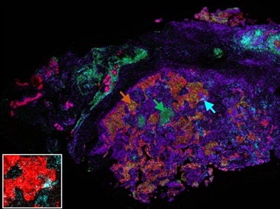
Multicolor MALDI-MS image overlay of breast cancer tissue section showing selected biomarkers, which highlight tumor-infiltrating immune cells. The orange, green, and cyan arrows indicate the tumor (co-localized CK and HER2), CD20+ B-cells, and CD8+ cytotoxic T-cells, respectively. The inset image on the lower left is a magnification of the region indicated by the cyan arrow, showing only CK and CD8. Credit: Highly Multiplexed Immunohistochemical MALDI-MS Imaging of Biomarkers in Tissues. Gargey Yagnik, Ziying Liu, Kenneth J. Rothschild, and Mark J. Lim Journal of the American Society for Mass Spectrometry 2021 32 (4), 977-988 DOI: 10.1021/jasms.0c00473
by Kenneth Rothschild, Ph,D, Chief Innovation Officer and Executive Chairman, Mark Lim, Ph.D, Executive Vice President and Chief Scientific Officer, and Gargey Yagnik, Ph.D, Principal Scientist and Director of Mass Spectrometry at AmberGen
Advancing the field of clinical research demands a holistic approach to mapping the spatial organization of molecules involved in biochemical pathways, an essential part of understanding disease pathology. However, the inherent limitations of traditional methods such as immunohistochemistry (IHC) mean only a few macromolecules of interest can be analyzed in a single scan. True multiomic imaging capability is emerging as the key to building a comprehensive and contextual understanding of the interactions of different biomolecules.
Developing a detailed understanding of the molecular landscape of health and disease requires more than the cataloging of individual biomolecules. The key challenge lies in mapping the spatial organization of proteins, nucleic acids, lipids and metabolites within the complex architecture of cells and tissues. Accurate contextualization of these molecular networks is essential in deepening our biological knowledge and uncovering mechanisms of disease.
Combining the technique of matrix-assisted laser desorption/ionization (MALDI) imaging, which has demonstrated robust untargeted analysis of small molecules, with new high-plex IHC technology capable of imaging large intact biomolecules such as proteins and nucleic acids in a targeted manner is enabling highly multiplexed protein detection on a single tissue section. This integration is breaking down longstanding barriers between metabolomics, lipidomics and proteomics interactions to support complex disease investigation.
The transformative effect of MALDI imaging
Since Richard Caprioli et al. first demonstrated MALDI mass spectrometry imaging (MALDI MS imaging) in the late 1990s, researchers have used the technique to visualize untargeted distributions of small biomolecules in intact tissue,1transforming studies of metabolites, lipids, glycans, extracellular matrix proteins and small drug compounds. The spatial resolution of larger macromolecules such as intact proteins and nucleic acids, however, was still limited. Methods such as IHC and in situ hybridization (ISH) provide targeted, high-resolution imaging of proteins and nucleic acids at cellular and subcellular levels, but are constrained by low multiplexing capability and the need for repeated, time-intensive cycles that can damage the tissue.
An integrated approach
Multiomics imaging is an important component of the fast-growing field of spatial omics, which is increasingly recognized as a method that provides the exact spatial coordinates of cellular and molecular profiles at the systemic level. The challenge here lies in combining these methods into a cohesive multiomic workflow capable of analyzing one tissue sample with a single instrument to offer a comprehensive biological picture.
The introduction of MALDI-IHC workflows that integrate antibody-based labeling with MALDI Imaging broke the mold. Early work using mass tag probes to detect nucleic acid in a complex mixture2 led to the realization that photocleavable linkers could be used to create photocleavable mass tagged (PC-MT) probes to image targeted proteins and nucleic acids in tissues. In the case of proteins, antibodies are conjugated with novel PC-MTs designed by AmberGen that act as molecular barcodes, which when detected by a MALDI mass spectrometer enable simultaneous imaging of hundreds of targeted intact proteins in a tissue sample.3,4
These PC-MTs are released from antibodies under controlled UV light exposure before MALDI analysis. Once detached, the well-defined mass-to-charge (m/z) ratios of the mass tag ions and absence of side products from the photocleavage reaction make them reproducible and easily detectable. These sharp, non-overlapping signal peaks reduce ambiguity in spectral interpretation and ensure that each target protein can be imaged simultaneously with high specificity.
This approach is compatible with standard histological workflows. Tissue staining with antibody-probe conjugates can be performed using conventional manual or automated immunohistology protocols and, after staining, controlled photocleavage of the probes liberates the tagged peptides prior to matrix application and MALDI imaging. Each liberated peptide provides a discrete, quantifiable signal, which can be reconstructed into a two-dimensional image showing the spatial distributions of multiple proteins. By layering these targeted protein maps with untargeted imaging of small molecules such as lipids, glycans, and metabolites, researchers can generate detailed molecular atlases that capture both the diversity and the spatial context of biological systems.
Uncovering insights into neurodegenerative disease
Alzheimer’s disease affects more than 55 million people globally5 and recent research combining MALDI imaging with PC-MT probes has shown promising results in gaining a better understanding of its progression3. In pharmaceutical research and development, characterizing the diverse biomolecular classes and cell types within the amyloid-β plaque microenvironment typical of Alzheimer’s disease remains a significant challenge. Simultaneous analysis of the complex interactions between proteins, lipids, and metabolites is required, as investigating any one of these biomolecule classes in isolation fails to capture its contextual interaction within the tissue. Ideally, multiple biomolecular classes would all be analyzed from a single tissue section using a benchtop imaging platform, although, until recently, integrating lipid and protein imaging on the same tissue slide has posed a significant technical challenge.
Recent advances in MALDI imaging are beginning to make this integration possible. An initial untargeted scan identifies label-free analytes such as lipids, metabolites and glycans while a subsequent run incorporates antibody-linked probes carrying PC-MTs to localize targeted proteins. This dual layer strategy enables the co-localization of small molecules with specific proteins in a truly multiomic workflow. Within the Alzheimer's model, this means proteins associated with plaque formation can be mapped, even in 3D, alongside surrounding lipids and metabolites to reveal how different biomolecular classes interact to drive pathology. MALDI-IHC is also helping to accelerate drug development for other neurodegenerative diseases such as Parkinson’s.6,7
Advances in studying lung cancer
Accurate lung cancer subtyping is critical to guide treatment decisions and monitor their effectiveness. Current methods that rely solely on traditional IHC are limited by the information that can be gained from a tissue sample. Complementing these processes with MALDI methods helps uncover more details of cancer subtypes.
To provide contextualization for tumor subtyping, MALDI-IHC was applied to a 99-core human lung tissue microarray (TMA) comprising normal, adjacent normal, adjacent cancerous, and cancerous tissues to determine molecular subtypes8. A 7-plex panel was initially used to identify subtypes in annotated tissues, while an expanded 23-plex panel was also applied to a 100-core human lung TMA. k-means clustering analysis demonstrated effective differentiation between subtypes.
Working toward achieving true multiomics
AmberGen, along with other researchers, are developing workflows that can capture a range of large and small molecules in layers within their spatial context to form a complete picture of their composition and location9. Recently this included imaging of mRNA transcripts.10 This is of clinical significance when studying heterogeneous tissues, such as cancer tumors where the ‘omic’ layers reveal the lipids, metabolites, drugs, proteins, and ribonucleic acid (RNA) from the same cell population if done on the same sample using the same instrument. The alternative would be to subject serial tissue sections to analysis using two different platforms, and therefore capture more than one cell population, which does not provide insight into tumor heterogeneity at the specific location.
In summary, the MALDI-IHC workflow for high plex spatial imaging brings the scientific community a step closer to achieving multiomic capability in research into diseases like cancer and Alzheimer’s. Integrating diverse molecular classes into unified spatial maps enables researchers to gain unprecedented insights into the interplay of cellular components. True multiomic workflows promise to transform the study of disease mechanisms and start to redefine the future of personalized therapies.