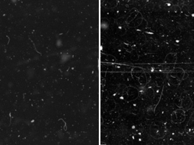
Glass coverslips (left) deliver consistent, high-quality imaging with minimal defects and a smooth, uniform surface, supporting reliable and reproducible results. Film coverslips (right), meanwhile, show nearly 5x more defects and increased surface irregularities.
by Cheryl Fudge, Senior Director, Global Product Management - Core Histology, Leica Biosystems
As pathology laboratories increasingly adopt digital workflows and artificial intelligence (AI)-powered diagnostic tools, the quality of every component in the imaging process matters more than ever. While scanners and software often receive the most attention, one often-overlooked element—the coverslip—may have a more significant impact on digital pathology performance than many realize.
When digital imaging and AI analysis depend on every pixel, "good enough" risks missed details and wasted time. As laboratories prepare for AI integration, understanding how different covering materials affect image quality is becoming increasingly important.
Why Coverslip Choice Matters for Digital Imaging
Cover glass and mounting media essentially form the initial optics of every biomedical microscope. Because modern microscope objectives achieve extremely high resolution, even slight variations in the optical properties of these components can influence the performance of the entire imaging system.
The most important factors to consider are how light passes through the material, the thickness of the covering, and how flat the surface remains.
When slides are scanned for digital pathology, the scanner's autofocus mechanism must work with whatever surface characteristics the coverslip presents. Materials that maintain consistent, predictable surfaces make this process more reliable, while irregular surface profiles may pose challenges for autofocus systems.
Surface Quality: The Foundation of Good Imaging
A recent comparative study conducted by a renowned optical research institute in Jena, Germany, examined the differences between conventional glass coverslips and polymer cover films.
One of the most important findings relates to their surface characteristics. Glass coverslips demonstrate exceptionally smooth surfaces—so smooth that in laboratory measurements, the roughness falls below what instruments can detect. Polymer films, by contrast, exhibit a characteristic uneven microstructure with measurable surface roughness of 5-6 nanometers.
At larger scales, both materials show some degree of surface unevenness when mounted to slides, but the patterns differ substantially. Glass coverslips display gentle, gradual curves with relatively wide spacing in the millimeter range. Polymer films show irregular rippling patterns with much tighter spacing of just a few hundred micrometers.
Perhaps most importantly for consistency, the study found that polymer film tends to follow the shape of the underlying tissue in its surface profile, while glass maintains its shape largely independent of the specimen beneath.
This means that with film, the surface characteristics can change depending on what tissue is being covered—introducing variability that digital systems must accommodate.
Impact on Image Quality
These surface differences translate into measurable effects on image quality and consistency. The study's optical simulations using scanner specifications showed that glass coverslip samples produce virtually no noticeable loss in optical performance, with contrast reduction below 0.1%.
There was no meaningful difference between blank samples and those with tissue sections.
Polymer film samples showed different results. Simulations demonstrated that image quality varies across different areas of the sample, with contrast dropping by 5-20% in some regions.
When testing how well each material preserves fine details using a digital resolution test pattern, glass maintained sharp resolution of 1.5 micrometers or better across the entire viewing area, while film samples showed decreased sharpness and areas where resolution dropped to greater than 2 micrometers.
The irregular surface patterns of polymer films can impair image quality at the limits of the microscope's resolution in certain areas of the sample.
While these effects occur primarily at resolution boundaries, they represent the kind of subtle variations that can compound when AI algorithms analyze images pixel by pixel.
Light Transmission and Optical Matching
Beyond surface texture, the study revealed that the two materials differ in how they handle light at a fundamental level. Glass coverslips are well-matched optically to standard microscope slides, which minimize image distortions.
Polymer film demonstrates a noticeably different refractive index—more than approximately 0.03 units different—meaning it doesn't match as well with the slide glass beneath it. This mismatch can result in slightly increased image distortions, particularly when using high-powered microscope settings.
Additionally, the study found that polymer film shows approximately 1-2% higher light loss compared to cover glass, likely due to light scattering on its rougher surface. At shorter wavelengths toward the blue end of the spectrum, these losses increase more dramatically, with absorption rising sharply below 400 nanometers.
Surface Defects: A Practical Concern
The study's assessment of surface defects revealed results with direct workflow implications. Automated inspection of blank samples showed that film-covered samples exhibit approximately five times more defects than glass-covered samples.
This difference exists even before any environmental exposure.
Analysis also identified lengthwise scratches present on film samples that were absent from glass samples. Since scanner resolution approaches 1 micrometer, defects in this size range can directly impact image quality. This is especially true for defects located between the covering material and the slide, where the scanner's very shallow focusing range of 1-2 micrometers makes them particularly problematic.
Interestingly, the study noted that film surfaces show less particle accumulation over time compared to glass when exposed to laboratory air. However, the initial defect count difference remains significant for laboratories concerned about baseline image quality.
Implications for AI-ready Workflows
These findings carry significant implications for laboratories implementing or planning AI-assisted diagnostic workflows. AI algorithms analyzing digital pathology images depend on consistent, high-quality input data. Cleaner scans with minimal artifacts reduce the need for time-consuming rescans while enabling more accurate AI analysis.
Surface artifacts, scratches, and optical inconsistencies introduce "noise" that can confuse automated analysis systems. When AI tools examine tissue at the pixel level, variations in image quality caused by coverslip irregularities may be misinterpreted or may obscure genuine features of interest. The approximately fivefold difference in surface defects between materials represents a substantial variable that laboratories should consider when standardizing their digital workflows.
Modern slide scanners rely on precise automatic focusing mechanisms. Glass maintains a highly stable and uniform surface that is crucial for consistent and reliable digital focusing, while the characteristic rippling pattern of film materials creates a more challenging focusing environment.
Practical Considerations
Laboratories evaluating their cover slipping practices for digital readiness should weigh these optical performance factors against their specific operational needs. As the study authors note, results reflect a single-site evaluation with specific instruments, protocols, and sample types, and outcomes may vary depending on laboratory environment, specimen characteristics, and workflow.
It is also important to note that the study assessed technical and optical performance only and did not evaluate clinical diagnostic accuracy or patient outcomes. The translation of optical quality differences into diagnostic impact would require separate clinical validation studies.
Small Choices Can Have Big Impacts
As digital pathology matures and AI-powered diagnostic tools become increasingly integrated into clinical workflows, attention to every element of the imaging chain becomes essential. The choice of coverslip material—often treated as an afterthought —can meaningfully influence the optical quality of scanned images.
Glass coverslips produce clean images with minimal artifacts, offering advantages in surface smoothness, optical consistency, and reduced surface defects that align well with the demands of digital imaging and AI analysis. For laboratories committed to optimizing their digital pathology infrastructure, understanding these material differences represents an important step toward ensuring the image quality that advanced analytical tools require.
The path to AI-ready digital pathology involves many decisions, from scanner selection to algorithm validation. Sometimes the most impactful choices are also the most fundamental—and the humble coverslip deserves more consideration than it typically receives.
About the author: Cheryl Fudge is Senior Director, Global Product Management - Core Histology at Leica Biosystems.