Microscope Image of Carnivorous Plant Wins 10th Anniversary Olympus BioScapes Competition
CENTER VALLEY, Pa., December 16, 2013 – Vegetarians beware! A spellbinding photo of the yawning trap of a carnivorous plant has taken First Prize in the 2013 Olympus BioScapes Digital Imaging Competition®, the world’s foremost forum for showcasing microscope images and movies of life science subjects. Igor Siwanowicz, a researcher from the Howard Hughes Medical Institute, Janelia Farm Research Campus, Ashburn, Va., captured the eerily fascinating photo of the floating humped bladderwort plant. The confocal image was selected from more than 2100 entries to earn Dr. Siwanowicz $5,000 worth of Olympus equipment. All of the award-winning BioScapes entries may be viewed online at www.OlympusBioScapes.com.

Celebrating its 10th Anniversary, the Olympus BioScapes Competition is the world’s premier platform for honoring images and movies of human, plant and animal subjects as captured through light microscopes. Images of any life science subject are eligible. Entries are judged based on the science they depict, their beauty or impact, and the technical expertise involved in capturing them. Entrants can use any brand of light microscope. In addition to the Top 10 award-winning recipients, 69 Honorable Mentions received recognition this year, including 55 still images and 14 movies.
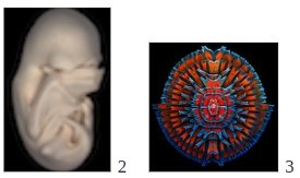
The 2013 winning entries reflect the latest advances in neuroscience and cell biology as documented by researchers, along with amazing glimpses of life on a microscopic scale captured by creative hobbyists, students and scientists. Specimens represent animal, plant and human subjects. Siwanowicz’s First Prize image is a good example. The aquatic carnivorous plant (Utricularia gibba) digests microinvertebrates sucked into its trap a millisecond after they touch its trigger hairs (hair bases are seen in the center of the image, inside the dome-shaped entrance). The traps also provide a microhabitat for single-cell green algae, predominantly desmids, which are visible in the photo. Siwanowicz also won Third Prize for his artful image of desmids, and earned an Honorable Mention for his image of rotifers surrounding a green alga. Second Prize went to a riveting image of a black mastiff bat embryo (Molossus rufus), at the "Peek-a-boo" stage, when its wings have grown to cover its eyes. The image was captured by Dorit Hockman, University of Oxford, Oxfordshire, UK.
This year’s honored images come from five continents, including 16 states of the U.S. and 22 other nations: Australia, Belgium, Brazil, Canada, Czech Republic, Denmark, England, Finland, France, Germany, Hungary, Iran, Italy, Japan, The Netherlands, Norway, Panama, Poland, Scotland, Spain, Switzerland and Wales. Competition participants hailed from a record 71 nations, making this year’s competition the most international to date.
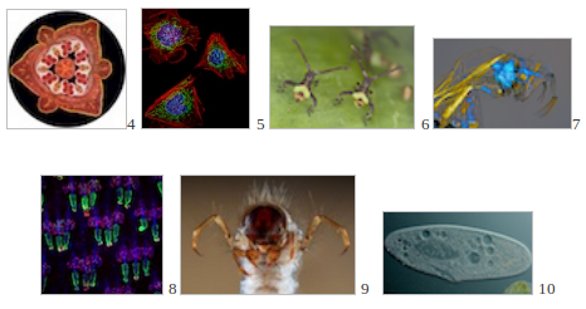
Subjects captured using wide variety of microscope imaging techniques dominated the Top 10 this year. Fourth Prize, a darkfield image of a lily flower bud, was captured by Spike Walker of Staffordshire, England. Dylan Burnette, of the National Institutes of Health in Bethesda, Md., earned Fifth Prize for a structured illumination microscopy fluorescence image of mouse fibroblasts. Kurt Wirz of Basel, Switzerland, earned Sixth Prize for a captivating image of a pair of “brother” insects, just two hours old. Charles Krebs of Issaquah, Wash., took Seventh Prize for a polarized light photo of the larva of a phantom midge. Yaron Fuchs, Howard Hughes Medical Institute/The Rockefeller University, New York, N.Y., captured 8th prize for confocal image of mouse tail showing stem cells, and Fabrice Parais of DREAL (Regional Directorate of Environment, Planning and Housing) in Basse-Normandie, Caen, France, won Ninth Prize for a stunning stereo microscope photo of the head of a caddisfly larva. Tenth Prize this year went to a fascinating video of a living Paramecium, captured using differential interference contrast by Ralph Grimm of Jimboomba, Australia.
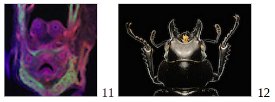
The winning images also show how visual themes sometimes repeat themselves in nature. For instance, two Honorable Mention images, one of mouse prostate and ureter by Xiaochen Lu and C. Chase Bolt of the University of Illinois at Urbana-Champaign, and another of a stag beetle captured by Viktor Sýkora of Hyskov, Czech Republic, bear a striking resemblance to each other.
“For 10 years, Olympus has sponsored this competition to bring to light the power, beauty and importance of science and the work that scientists do,” said Brad Burklow, Executive Director of International Business for the Scientific Equipment Group of Olympus America Inc. “BioScapes movies and still images combine art and science to remind us of the fascination and wonder of the natural world and highlight vital work going on in laboratories. They serve as an inspiration to young people seeking careers in science, technology, engineering and mathematics. For a decade, Olympus has been dedicated to bringing these amazing images and stories to the attention of scientists and non-scientists worldwide.”
The winners were announced last night at a gala reception in New Orleans, marking the 10th Anniversary of the competition. A selection of the 2013 winning and Honorable Mention images and videos will be displayed in a museum tour that will travel the U.S. over the coming year. The 2013 winners’ tour of BioScapes images is sponsored by Olympus America in cooperation with Scientific American. Other exhibits of winning BioScapes images also will tour cities across the U.S., Mexico, South America, Canada and the Middle East throughout 2014.
Olympus selects outstanding authorities in microscope imaging as judges for each year’s competition. This year’s BioScapes panel of judges included the eminent James E. Bear, Ph.D., University of North Carolina-Chapel Hill; Brian Matsumoto, Ph.D., University of California, Santa Barbara (emeritus); Alison North, Ph.D., The Rockefeller University, New York; and Lei Zhu, Ph.D., Florida State University.
Next year’s competition, which closes September 30, 2014 is already open for participants. Entrants can submit up to five still images, image sequences, or movies of life science subjects captured at any magnification using a compound light microscope. The judges make their decisions without participant or brand information. To enter the competition or view the BioScapes gallery of winners and Honorable Mentions, visit www.OlympusBioScapes.com. For free access to the images, media members and other noncommercial users may contact contact [email protected]
About Olympus
Olympus is a global technology leader, designing and delivering innovative imaging solutions in its core business areas: Medical and Surgical Products; Life Science Imaging Systems; Industrial Measurement and Imaging Instruments; and Cameras and Audio Products. Through this technology, Olympus focuses on enhancing people’s lives every day and helping people enjoy the continuum of life. For more information, visit www.olympusamerica.com.