Opterra Offers Superior Integration of Confocal Microscopy and Photoactivation for Biology Applications
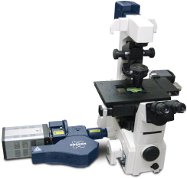
NEW ORLEANS, Louisiana – December 16, 2013 – Today at the 2013 American Society for Cell Biology Annual Meeting, Bruker introduced the [1]Opterra Multipoint Scanning Confocal Microscope, which sets a new standard for integration of confocal imaging with photoactivation. The new Opterra microscope utilizes a number of innovative features to obtain the speed of wide-field imaging and the resolution of traditional confocal systems while minimizing phototoxicity, making it an ideal solution for gentle and fast confocal imaging of live cell preparations. A seven-position pinhole/slit aperture allows the Opterra to be optimized for varying objective lens magnifications that results in the ability to image deeper into tissue versus conventional disk scanning confocal microscopes.
“The Opterra has proven to be a major advance in terms of rapid, time-based volumetric imaging,” said Dr. Mario De Bono, Medical Research Council Group Leader at the Laboratory of Molecular Biology, Cambridge University, UK. “The speed of the system, coupled with its sensitivity and resolution has significantly enhanced our ability to visualize neural activity in 3D in C. elegans at speeds that were previously not possible. The ability to change pinhole size is great, as it allows us to match the imaging setup with the specimen.”
“Our new Opterra provides a flexible optical workstation for cell biologists to perform confocal imaging of live cells and small organisms with simultaneous point and area scanning for photoactivation and photoablation,” explained Mike Szulczewski, Vice President and General Manager of Bruker's Fluorescence Microscopy business. “The tight integration of optical imaging with optical stimulation techniques enables investigators to take full advantage of today’s imaging and photochemical probe technologies.”
About Opterra
The Opterra Multipoint Scanning Confocal Microscope is based on Bruker’s patented swept-field imaging scanner. This scanner allows high-speed confocal imaging of live cell and small organism preparations at resolutions comparable to conventional point scanners, but with minimal phototoxicty. Opterra includes a second scanner for photo- activation/bleaching/ablation, which can operate simultaneously with imaging. The photoactivation scanner can be coupled to both visible and multiphoton lasers, thus allowing the use of the full range of photo-activatable molecules and photochemical techniques available to life science researchers. In the case of multiphoton lasers, this provides precise three-dimensional control over photoactivation. The applications addressed by Opterra include response to DNA damage, kinetics of photoactivatable fluorescent proteins, fluorescence recovery after photo-bleaching (FRAP), response to local stimulation of channel proteins, and response to cell membrane damage. Bruker’s Prairie View 5.0 software provides an intuitive interface with a rich environment for defining image acquisition and photoactivation protocols.
About Bruker Corporation (NASDAQ: BRKR)
Bruker Corporation is a leading provider of high-performance scientific instruments and solutions for molecular and materials research, as well as for industrial and applied analysis. For more information about Bruker Corporation, visit www.bruker.com.