MIT engineers have created a new diagnostic nanoparticle that combines imaging technologies and molecular diagnostics. In a new study, Sangeeta Bhatia and her colleagues showed that the diagnostic could be used to monitor the progression of colon cancer, including the spread of metastatic tumors to the lung and the liver. Eventually, they hope it could be developed into a routine cancer test that could be performed annually.
The researchers wanted to develop what they call a "multimodal" diagnostic, which can perform both molecular screening (detecting the urinary signal) and imaging, to tell them exactly where the original tumor and any metastases are located. To modify the particles so they could also be used for PET imaging, the researchers added a radioactive tracer called copper-64. They also coated them with a peptide that is attracted to acidic environments, such as the microenvironment in tumors, to induce the particles to accumulate at tumor sites. Once they reach a tumor, these peptides insert themselves into cell membranes, creating a strong imaging signal above background noise.
The researchers tested the diagnostic particles in two mouse models of metastatic colon cancer, in which tumor cells travel to and grow in the liver or the lungs. After treatment with a chemotherapy drug commonly used to treat colon cancer, the researchers were able to use both the urine signal and the imaging agent to track how the tumors responded to treatment.
The researchers also found that delivering copper-64 with their nanoparticles offers an advantage over the strategy that is typically used for PET imaging. The PET tracer, known as FDG, is a radioactive form of glucose that is taken up by metabolically active cells, including cancer cells. However, the heart generates a bright PET signal when exposed to FDG, and that signal can obscure weaker signals from nearby lung tumors. Using acid-sensitive nanoparticles to accumulate Copper-64 in the tumor environment provides a much clearer image of lung tumors, the researchers found.
If approved for use in human patients, Bhatia envisions that this kind of diagnostic could be useful for evaluating how well patients respond to treatment, and for long-term monitoring of tumor recurrence or metastasis, especially for colon cancer. In the longer term, she hopes that this technology could be used as part of a diagnostic workflow that could be given periodically to detect any kind of cancer.
"The vision is that you could use this in a screening paradigm—alone or in conjunction with other tests—and we could collectively reach patients that do not have access to costly screening infrastructure today," she says. "Every year you could get a urine test as part of a general check-up. You would do an imaging study only if the urine test turns positive to then find out where the signal is coming from. We have a lot more work to do on the science to get there, but that's where we would like to go in the long run."
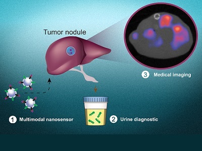
Photo: Multimodal nanosensors (1) are engineered to target and respond to hallmarks in the tumor microenvironment. The nanosensors provide both a noninvasive urinary monitoring tool (2) and an on-demand medical imaging agent (3) to localize tumor metastasis and assess response to therapy. Credit: Liangliang Hao