Scientists have made strides in understanding the structure and building blocks of genes and RNA with the help of methods like next-generation sequencing (NGS), single-cell RNA sequencing (scRNA-seq) and polymerase chain reaction (PCR) amplification. However, the proteins that are built from these genetic blueprints cannot be amplified and sequenced in the same way, and fully understanding how and where proteins work within the body has been relatively more difficult. Researchers from Northwestern University and the University of Pittsburgh have taken a step toward better mapping the proteomes within living cells with a new technique that targets proteins in the neurons of live mouse brains.
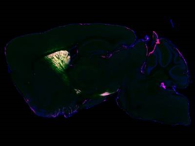
The researchers designed a virus to tag proteins with APEX2 for proximity labeling, specifically targeting neurons located in the striatum of the living mouse. An enhanced green fluorescent protein (EGFP) was used to show which neurons APEX2 was expressed in. After validating the technique with fluorescence and electron microscopy, the researchers found the technique provided a snapshot of the entire proteome inside living neurons, which can be analyzed postmortem with mass spectrometry. The study was published in Nature Communications.
“Similar work has been done before in cellular cultures. But cells in a dish do not work the same way they do in a brain, and they don’t have the same proteins in the same places doing the same things,” said senior author Yevgenia Kozorovitskiy, of Northwestern University. “It’s a lot more challenging to do this work in the complex tissue of a mouse brain. Now we can take that proteomics prowess and put it into more realistic neural circuits with excellent genetic traction.”
“Mass spectroscopy-based proteomics is a powerful technique,” said first author Vasin Dumrongprechachan, a Ph.D. candidate in Kozorovitskiy’s laboratory. “With our approach, we can start mapping the proteome of various brain circuits with high precision and specificity. We can even quantify them to see how many proteins are present in different parts of neurons and the brain.”
Now that the system is validated, the researchers can apply it to more mouse models to better understand neurological diseases. Dumrongprechachan said the approach could help identify neuronal protein changes that occur during specific patterns of brain activity or in response to neuroactive drugs. And by applying the technique to mouse models related to brain diseases, the researchers can make connections to postmortem proteomics work involving the human brain, according to Kozorovitskiy.
Photo: Protein expression captured in the mouse brain, visualized by fluorescence microscopy. Image shows a sagittal cross section of the mouse striatum. Blue is the outline of the brain. Green and magenta show selectively tagged proteins for mass spectrometry analysis. Credit: Northwestern University