The ability to visualize the 3D distribution of specific cell types in a whole organ can help scientists better understand the differences that arise due to disease, and ultimately come up with better treatments and diagnostic methods for conditions like type 2 diabetes and cancer. While methods for 3D imaging large samples such as entire human organs have advanced in recent years, highly specific labeling of certain cell types, such as insulin-secreting islets of Langerhans in the pancreas, remains difficult within large tissue volumes. Researchers at Umeå University have presented a potential solution for specific labeling and high-resolution 3D imaging of entire organs, using a 3D-printed matrix and computational reconstruction techniques.
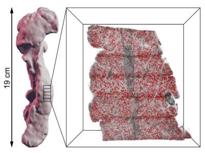
The researchers demonstrated their new method by imaging and mapping the distribution of islets within the pancreata of both non-diabetic donors and donors with type 2 diabetes. The 3D-printed matrix was designed to provide optimally-sized slices of organ for fluorescent labeling, optical projection tomography and light sheet fluorescence microscopy. The matrix also provides a template for specific coordinates to be assigned to each slice, allowing for precise 3D reconstruction of each portion within its original spatial context.
The method resulted in micrometer-resolution 3D images of the pancreata, providing a detailed map of the labeled islets throughout the whole organ. The researchers were able to demonstrate previously unrecognized features of pancreatic anatomy, including areas of extremely high islet density in both the diabetic and non-diabetic individuals. The study was published in Communications Biology.
“Besides using the new method to study diabetes, it can also improve understanding of other pancreatic diseases, not least pancreatic cancers, and we have initiated collaborations with clinical researchers in Umeå to look into that,” said corresponding author Ulf Ahlgren. “But the technology itself should be possible to use to study other organs and diseases in similar ways since it enables the study of where cellular changes take place in a full organ context, their amount and relationship to nearby tissues and cell types.”
The new method may contribute to an advanced understanding of the cellular changes related to different diseases, Ahlgren said. Further study of the pancreas using this technology could lead to improved islet transplantation protocols for diabetic patients and the development of non-invasive clinical imaging strategies.
Photo: Illustration of how a pancreas can be divided into smaller parts that are labelled depending on their specific cell types using anti-insulin antibodies. The parts can then be pieced together using a computer as a three-dimensional jigsaw puzzle to recreate the entire human organ. Credit: Max Hahn and Ulf Ahlgren