Advancements in optical microscopy have made it possible to view large biological samples in cellular or even subcellular resolution, but doing so requires a large amount of light, which can interfere with fluorophores and cells. Scanning and irradiating an entire sample can lead to photobleaching or alterations in cell physiology, but reducing the amount of light through the whole sample also reduces resolution. Researchers from the Institut Fresnel at Aix Marseille University have proposed a new scanning fluorescence microscopy method that significantly decreases the amount of light sent into the sample, without sacrificing resolution, using algorithms that allow the microscope to adapt to the sample’s architecture.
The new adaptive technique is based around the fact that most biological tissues have a well-known architecture, such as epithelia found in embryos, which are made up of curved cell sheet surfaces. The researchers designed an algorithm that incorporates this knowledge to allow the microscope to predict the surface structure based on data from a preliminary scan, and then tailor its scanning pattern to the estimated morphology. This enables the microscope to target specific areas along the surfaces of interest and direct light to those areas rather than irradiating the entire sample.
The smart microscope used to examine live Drosophila melanogaster epithelia and produced high-resolution images while using up to 80 times less light than a typical full scan. The technique can also be easily implemented on most commercially available scanning fluorescent microscopes, the researchers said. This study was published in Light: Science & Applications.
The adaptive microscopy method opens up new opportunities for imaging live biological samples like embryos and organoids over long periods of time without interfering with their physiology.
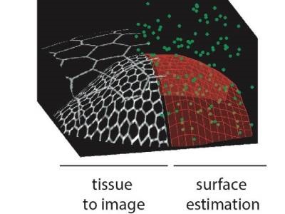
Photo: (Left) A drawing of curved biological tissue. The hexagons represent the fluorescent contours of the cells organized in a cell sheet. The tissue can be covered by a second epithelium, which can be ignored in the imaging process. (Right) From a few acquisitions (green dots), the microscope automatically estimates the surface of the tissue (red mesh) and can then concentrate the acquisitions on this surface, or even only on the fluorescent cell contours thanks to a propagative acquisitions algorithm. Credit: Faris Abouakil et al.