Understanding where drug molecules bind to their targets is an essential element of drug development, and drug-target interaction studies help predict both the therapeutic effects and side effects of a drug. However, precise observation of drug-target interactions in situ is difficult, in part due to the diffusion or blocking of fluorescence by intact animal tissue, and these studies may rely on imprecise methods such as bulk analyses of drug-molecule concentration in entire organs. A new technique developed at Scripps Research overcomes some of the challenges of in situ drug-target identification by combining tissue clearing and click chemistry to precisely image labeled molecules with subcellular resolution.
The technique, called clearing-assisted tissue click chemistry (CATCH), enabled the researchers to perform brain-wide imaging of covalent drug-target engagement in intact mouse brain tissue. Following the injection of drug molecules containing probes, the tissue is cleared and fluorescent labels are affixed through a specific click reaction with the probes, allowing the drug-target engagement sites to be clearly visualized. Immunolabeling also allows specific cell types and subcellular compartments targeted by the drug to be identified.
The researchers tested several different fatty acid amide hydrolase (FAAH) inhibitors in their study, and found they could image the drug-target interactions of these inhibitors at the single-cell level within large volumes of mouse brain tissue. Different patterns of target engagement for each drug could be easily distinguished using the CATCH method. Among the FAAH inhibitors study was BIA-10-2474, which caused one death and several injuries in a clinical trial in 2016; the team found the inhibitor engaged unknown targets in the midbrain of mice, even when the mice lack the FAAH enzyme, offering a potential clue to the source of the drug’s toxicity.
Combined with immunolabeling, the CATCH method enabled identification of target cell types, including rare cell types, and allowed researchers to distinguish engagement sites in different parts of neurons. The method also revealed dose-dependent target shifts across different tissue, cellular, and subcellular compartments, which cannot be distinguished through conventional methods. This research was published in the journal Cell.
“This method ultimately should allow us, for the first time, to see relatively easily why one drug is more potent than another, or why one has a particular side effect while another one doesn’t,” said senior author Li Ye. “... The unique environment at Scripps Research, where biologists routinely work together with chemists, is what made the development of this technique possible.”
The team plans to further develop CATCH for use on thicker tissue samples, which ultimately could include whole mice. They also hope to extend the approach to non-covalently-binding drugs and chemical probes, and believe the method could become a basic tool not only for drug discovery, but other areas of biological research.
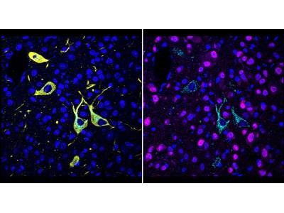
Photo: A team at Scripps Research invented a new method, called CATCH, that shows how drugs hit their targets in the body. Cells targeted by a drug (pargyline shown in cyan) can be identified by multiple rounds of immunolabeling (red showing neurons; yellow showing dopaminergic/noradrenergic neurons; blue showing cell nuclei).