Human organoids are valuable models for biomedical researchers to study tissue development, drug interactions and other biochemical functions of tissues and cells. Detailed 3D images of these unique cultures help scientists observe changes in response to different conditions, but conventional scanning-based imaging techniques have certain drawbacks such as being time consuming, having limited temporal resolution and potentially subjecting samples to photodamage. Researchers at the Georgia Institute of Technology (Georgia Tech) have now built a custom microscope system designed to overcome some of these challenges and capture 3D images of human organoids at cellular resolution in just milliseconds.
The team’s system was built “from scratch on an optical table,” according to Coulter BME Assistant Professor Shu Jia, who led the research, and was specifically designed for imaging “at the tissue and animal scale.” The system is based on Fourier light-field imaging and employs a hybrid point-spread function to produce highly-detailed reconstructions of the 3D volume of tissues from a single capture. Because the method, abbreviated as hPSF-FLFM, forgoes time-consuming scans, it allows for dynamic processes to be captured and characterized with greater spatiotemporal resolution and also serves to minimize photodamage.
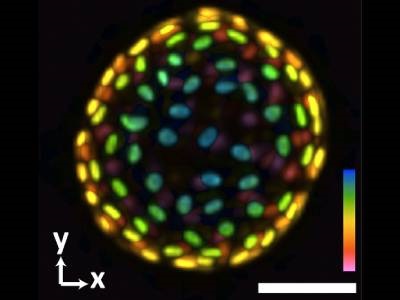
The Jia lab researchers demonstrated the capabilities of the hPSF-FLFM system by imaging the responses of human induced pluripotent stem cells-derived colon organoids (hCOs) to extracellular cues such as osmotic and mechanical stresses. The system allowed the team to observe dynamic changes at the cellular level across the entire organoid in 3D, with each raw image captured in approximately 0.1 seconds. The results showed the promise of hPSF-FLFM as an avenue to further improve organoid research, the authors concluded. This study was published in Biosensors and Bioelectronics.
“Because this is a custom-built system, it’s very flexible and adaptive,” said Jia. “It works with organoids, but similarly, it can work with animal models. I think we can extend this method to different areas of research. There are a number of potential collaborations we are exploring.”
The hPSF-FLFM system builds on the Jia lab’s ongoing work focused on next-generation imaging systems. This latest research was supported by funding from the National Institutes of Health and National Science Foundation.
Photo: An image of a colon organoid captured by a new system developed in Shu Jia's lab that can capture dynamic, 3D information about the lab-grown cultures of tissues in a single image. The raw image was captured in 0.1 seconds. The coloration represents depths of 60 and -60 micrometers from the focal plane to depict the organoid in three dimensions. Credit: Georgia Tech