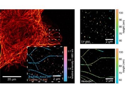
MIET-SMLM imaging of microtubules and clathrin pits in COS7 cells. Spectral splitting (inset) allows to effi-ciently distinguish between two different target and MIET imaging (right panel) gives height profiles with exceptionally high resolution. Credit: University of Göttingen
Super resolution imaging techniques have provided great advances in the observation of nanoscopic cell structures, however, a large gap still exists between lateral and axial resolution. This gap, in which axial resolution falls behind lateral resolution by a factor of two to three, limits the ability to view biological structures as they truly exist – in 3D. A research team led by Göttingen University, including the University of Würzburg and the Center for Cancer Research in the US, recently combined two imaging techniques, allowing them to achieve isotropic super resolution for imaging of biological samples.
The team combined two techniques that can achieve resolution in the nanometer range – metal-induced energy transfer (MIET) and direct stochastic optical reconstruction microscopy (dSTORM). The exceptional depth resolution of MIET imaging, combined with the extraordinary lateral resolution of dSTORM, helped bridge the gap in 3D super resolution imaging to achieve isotropic resolution. In addition, the researchers implemented dual-color MIET-dSTORM, enabling them to image two different cellular structures in three dimensions at once.
The researchers demonstrated their method by imaging U20S cells with microtubules and clathrin coated pits labeled with different dyes. MIET-dSTORM was performed using a confocal microscope and the cells were seeded on a cover glass coated with 10 nm of gold and 3 nm of SiO2 using standard immunofluorescence sample preparation procedures. The technique was shown to be compatible with biological samples and enabled high localization procession in both the xy and z directions, enabling nanoscopic resolution in all three dimensions. This research was published in Science Advances.
“The beauty of the technique is its simplicity. This means that researchers around the world will be able to implement the technology into their microscopes quickly,” said Jörg Enderlein, who led the research team at the Biophysics Institute, Göttingen University.
MIET-dSTORM offers a powerful tool for resolving protein complexes and small organelles with sub-nanometer accuracy, said corresponding author Oleksii Nevskyi, adding that anyone with access to confocal microscope technology with a fast laser scanner and fluorescence lifetime measurement capabilities can try the technique.