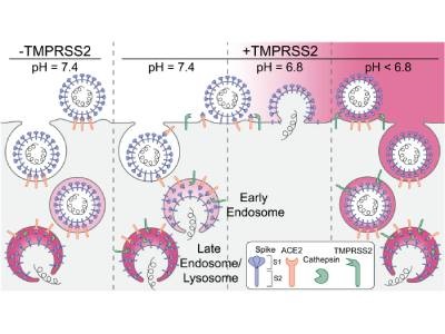
Schematic representation of the principal entry routes SARS-CoV-2 uses for infection. Entry starts with membrane attachment and ends with S protein–catalyzed membrane fusion releasing the viral contents into the cytosol. Fusion activity depends on two proteolytic cleavage steps, namely, one typically carried out by furin in the producing cell and the second by TMPRSS2 on the cell surface on in endosomes of the target cell. Alternatively, endosomal cathepsins can carry out both cleavages. Exposure of the virus to an acidic milieu is essential for membrane fusion, genome penetration, and productive infection. Fusion and penetration occur only in acidic early and late endosomal/lysosomal compartments but not at the cell surface, even when the furin and TMPRSS2 cleavages have both occurred. Fusion and penetration can occur at the cell surface of cells expressing TMPRSS2 if the extracellular pH is ∼6.8. Credit: Proceedings of the National Academy of Sciences (2022). DOI: 10.1073/pnas.2209514119
Understanding the fundamental mechanisms of viral infection could lead to new methods for intervening before the onset of infections like COVID-19. Recently, researchers have used genetic engineering and high-resolution microscopy techniques to visualize these tiny microbes in action, gaining a better understanding of each step in the process of virus entry. A team led by researchers from Harvard Medical School, Boston Children’s Hospital, Washington University and the University of Helsinki developed an approach to track single virions in real-time and found that SARS-CoV-2 requires acidic pH conditions to successfully infect cells.
The researchers captured live footage of the virions as they made entry into cells using lattice light sheet microscopy, along with chemical and genetic engineering methods. The team engineered a chimeric vesicular stomatitis virus to include the SARS-CoV-2 spike protein, which allowed them to separately label and visualize the spike protein and viral genome. The researchers captured a video covering four minutes of activity in real time and in three dimensions with snapshots taken every four seconds. The first part of the video below shows the engineered virus with the spike protein (labeled pink) as it is captured at a cell surface and engulfed by an endosome; the virus then fuses with the endosome membrane and injects its genetic material (labeled blue) inside the cell. The second part of the video shows many such viruses inside the cell.
Video Credit: Harvard Medical School
Observing each step in the entry pathway under different pH conditions allowed the researchers to determine the pH conditions required for infection and provided insights into the mechanisms through which pH impacts infection. The team found that the viruses cannot fuse with the membrane and release their genomes unless the environment is slightly acidic, with a pH between 6.2 and 6.8. Their observations showed that the acidic environment allows enzymes in the endosome or on the cell surface – including TMPRSS2, a key enabler of SARS-CoV-2 infection – to cut the spike protein and facilitate membrane fusion. Testing healthy volunteers using a pH catheter, the researchers further found that the human nose, where SARS-CoV-2 infection often begins, has an average pH of about 6.6. This research was published in the Proceedings of the National Academy of Sciences.
“Amusingly enough, measuring the pH of the nostril cavity has rarely been done before,” noted co-senior author Tomas Kirchhausen, professor of cell biology in the Blavatnik Institute at Harvard Medical School and professor of pediatrics at Boston Children’s Hospital.