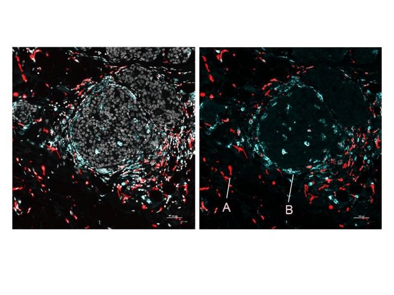
Image of human breast cancer cells showing A) immunosuppressive macrophages near tumor connective tissue, and B) immunostimulatory macrophages near tumor nests. Credit: Nir Ben Chetrit
Transciptomics and proteomics analysis techniques can reveal key information about the activity of cells, with RNA and proteins providing complementary data toward a better understanding of biological processes. However, many high-resolution transcriptomics and proteomics analysis methods require the dissolution of tissues, and thus the loss of spatial information, while many techniques that maintain spatial data lack the capability to probe both RNA and proteins simultaneously. Researchers at Weill Cornell Medicine, NewYork-Presbyterian and the New York Genome Center, in collaboration with biotechnology company 10x Genomics, have now developed a method that integrates spatial transcriptomics and proteomics, using unique probes to track the location and identity of both messenger RNAs (mRNAs) and proteins.
The new technique, called Spatial PrOtein and Transcriptome Sequencing (SPOTS), is based on existing technology from 10x Genomics that uses glass slides coated with special probe molecules to enable spatial transcriptomics. The probes, which contain molecular “barcodes” indicating their x and y position on the slide, bind to adjacent cells’ mRNAs when a thinly sliced tissue sample is made permeable and placed on the side. In addition, the method uses designer antibodies to bind to specific proteins of interest, which also bind to the probe molecules, providing both proteomic and spatial information. The method allows the mRNAs and selected proteins to be mapped simultaneously and identified using automated techniques, quickly unveiling a wealth of complementary data about tissue architecture and organization, cell activities and cell-to-cell interactions, according to co-senior author Dan Landau of Weill Cornell Medicine, NewYork-Presbyterian and the New York Genome Center.
The team first demonstrated SPOTS using healthy mouse spleen tissue, unveiling the complex functional architecture of the organ, including clusters of different cell types, their functional states and how these states varied with cell location. Additionally, the researchers used SPOTS to map the cellular organization of a mouse breast tumor. The map revealed macrophages in two distinct states, one fighting the tumor and the other immune-suppressive and forming a barrier to protect the tumor. This analysis highlighted SPOTS’ potential use in cancer research, with the macrophage subsets found in different areas of the tumor and interacting with different cells, their states likely driven by differences in the tumor microenvironment, according to Landau. These details could help explain why some patients respond to immune-boosting therapy while others don’t, and could inform the development of future immunotherapies, Landau added. This research was published in Nature Biotechnology.
“The initial version of SPOTS has a spatial resolution such that each ‘pixel’ of the resulting dataset sums gene activity information for at least several cells. However, the researchers hope soon to narrow this resolution to single cells, while adding other layers of key cellular information,” Landau said.