Polystyrene nanoparticles cause cardiac malformations by disrupting the development of the cardiac neural crest. Credit: Environmental International (2023). DOI: 10.1016/j.envint.2023.107865
Microplastics and nanoplastics pose serious concerns for both human health and the health of the environment and natural ecosystems. With recent studies revealing the presence of microplastics in lung tissue and blood from living humans, more research is needed to examine the potential health and developmental impacts of this pollution. Researchers at the Institute of Biology Leiden at Leiden University recently conducted a study using chicken embryos which revealed how nanoplastic particles can target embryonic stem cells and cause a number of severe malformations in the developing vertebrates.
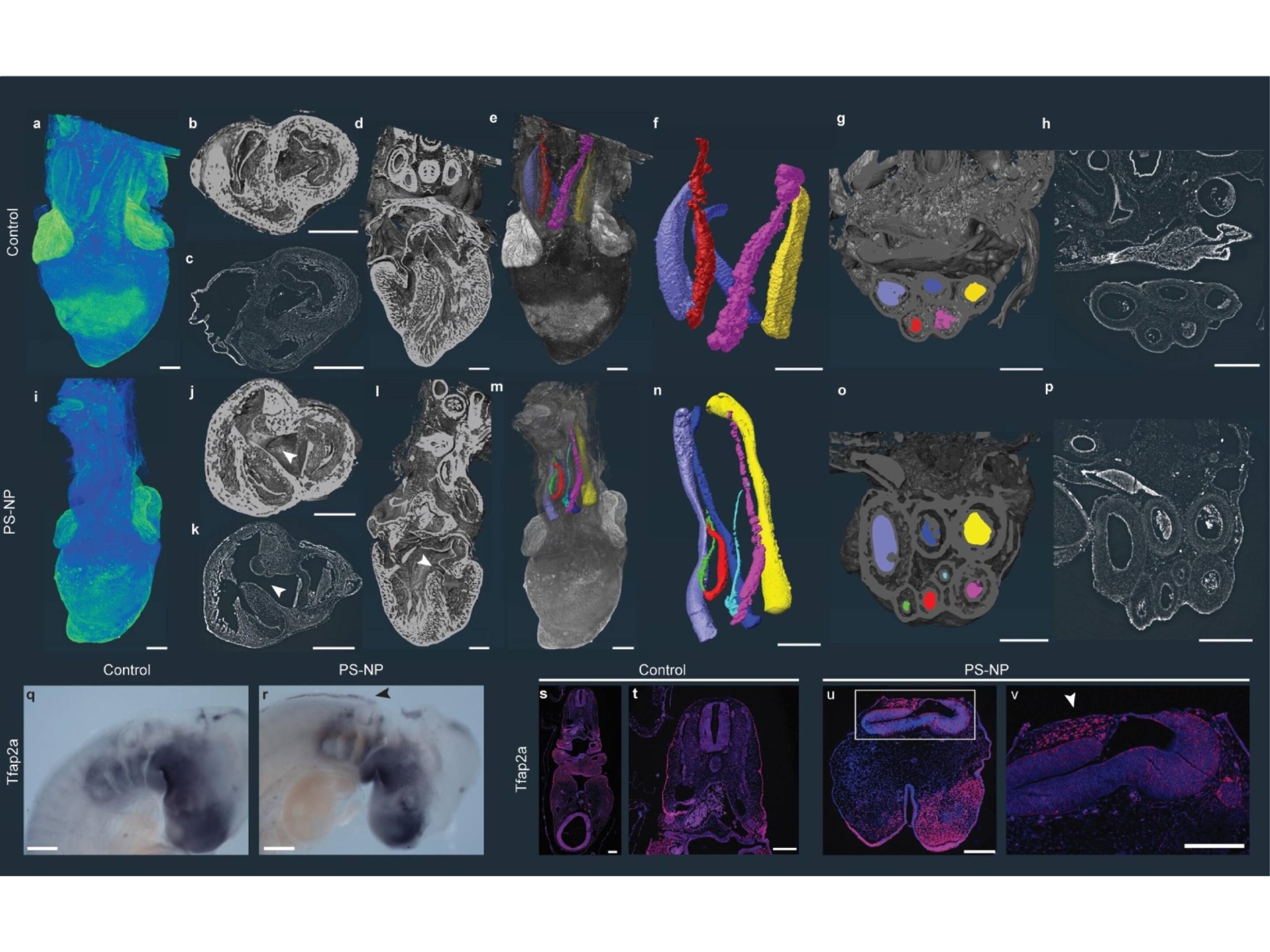
The researchers exposed the embryos and embryo-derived cell cultures to polystyrene particles ranging in size from 25 nm to 1 µm, including fluorescent particles to aid in visualization. In addition to confocal microscopy, the researchers also utilized X-ray microtomography (microCT), synchrotron X-ray tomographic microscopy and transmission electron microscopy (TEM) to visualize the development of the embryos and the distribution of plastic nanoparticles in the cultures and embryos. MicroCT scanning enabled detailed 3D reconstructions of the embryos to be studied for malformations, and synchrotron X-ray tomographic microscopy provided a high resolution view of the impact of nanoplastics on the heart and arteries.
The experiments revealed how the plastic particles passively target neural crest cells, resulting in developmental disruptions and malformations in multiple areas. The results suggest that the polystyrene particles selectively bind to the neural crest cells, preventing their migration or causing cell death. Because the migration of neural crest cells plays an important role in the development of multiple organs and structures in vertebrates, the chicken embryos developed with several malformations, including defects of the heart, eyes and face. This study, the first to reveal the mechanisms through which nanoplastics can disrupt these embryonic cells, was published in Environment International.
“We used a high concentration of polystyrene particles, that would normally not be present in an organism. But it shows what nanoplastics can do in extreme cases on very young embryos,” noted first author Meiru Wang. “And it also gives us guidelines on what can happen less severely in the developmental stage.”
The study is also the first to provide evidence that nanoplastics can lead to serious defects in the heart and great vessels, insights made possible by the Swiss Light Source synchrotron at the Paul Scherrer Institut. MicroCT experiments were performed at the Naturalis Biodiversity Center in Leiden, and additional collaborators include the Institute of Psychology, Methodology and Statistics at Leiden and Universitas Gadjah Mada in Indonesia.