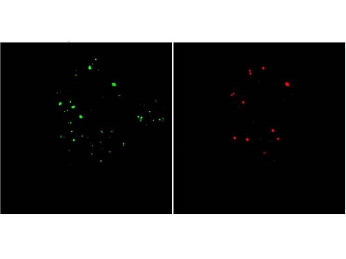
A HeLa cell labeled by the new probe shows total endolysosomes in green, with late endosomes/lysosomes in red. Credit: Jiajie Diao
Lysosomes are vital organelles that break down and recycle waste within the cell; when lysosomes do not function properly, waste can build up and become toxic to the cell. Endolysosomes are lysosomes that begin as endosomes, but the conversion of endosomes into lysosomes is difficult to study in detail. Researchers at the University of Cincinnati (UC) have developed a new fluorescent probe that can enable the tracking of this conversion process through a pH-sensitive color change.
When endosomes transform into endolysosomes, their pH levels change from a neutral to an acidic environment; an acidic environment is required for lysosomes to function properly. UC researchers previously worked on a pH-sensitive probe called ECGreen that turns brighter in a more acidic environment, but this change in brightness only provides relative information that cannot be precisely quantified. The new technology, a cationic quinolinium-based fluorescent probe called PyQPMe, instead shifts from green to red from a neutral to acidic environment. The red and green signals provide more detailed and precise information about the pH environment, enabling the researchers to better monitor the transition from endosomes to endolysosomes.
The team used the PyQPMe probe to observe live cells and found that the probe’s high fluorescence intensity reduced background noise, providing high resolution. The researchers were able to precisely measure the transition from early endosomes, to late endosomes, to endolysosomes, finding that a consistent ratio of endosomes were converted into lysosomes under various conditions. The conversion rate was constant in different environments and between normal and abnormal cells, a discovery that can aid in a better understanding of lysosomal diseases. For example, the cell may not be able to increase the number of lysosomes available in a situation where it is needed, such as when a cell is damaged. The probe technology could further be used to gather more information about endolysosomes, such as their location, number and size, which could ultimately be applied to the clinical diagnosis of lysosomal disease risk, explained co-lead author Jiajie Diao. This research was published in ACS Sensors.
“Our research results demonstrate that it is an extremely promising direction in designing and developing small fluorescent probes for bioimaging applications, especially when coupled with high-resolution microscopy,” said co-lead author Yujie Sun. “We will continue developing novel fluorescent probes with multiple functions.”
The researchers are also developing a machine-learning software that could be used to profile cell abnormalities and quickly diagnose lysosome problems based on information from the color-changing probe.