Credit: Tsukada et al.
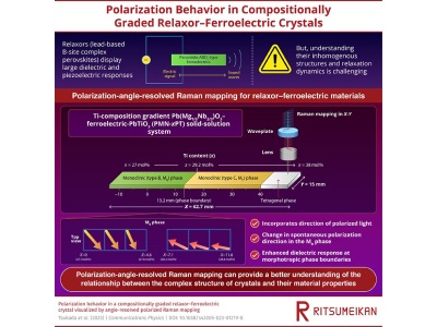
Researchers from Ritsumeikan University in Japan have developed a polarization-angle-resolved Raman microscope to study the arrangement and fluctuation of electric polarization in ferroelectric materials. By combining a half-wave plate with a Raman microscope, researchers were able to create a methodology for identifying vibration states and directions in a material.
In the study, published in Communications Physics, the team applied the newly developed methodology to investigate a piezoelectric lead magnesium niobate-lead titanate crystal commonly used in ultrasound equipment. These PMN-PT crystals have distinct dielectric and piezoelectric responses at their phase boundaries. By utilizing the new technique the researchers were able to map the titanium ratios which create these phases and phase boundaries. What the spectra showed was a slower relaxation of polarization at certain phase boundaries and they found that a slow response enables the material to store a large amount of charge, leading to an enhanced dielectric response.
“The development of this polarization-angle-resolved Raman microscope along with the advancements in analytical techniques can enable the incorporation of polarization information into the existing Raman imaging data and allow a deeper understanding of material properties,” said Professor Yasuhiro Fujii.
The implementation of the newly developed polarization-angle-resolved microscope could be used to optimize materials in the future including optimizing their dielectric performance. This could lead to improvements in ultrasound technologies and improved ultrasound detection.