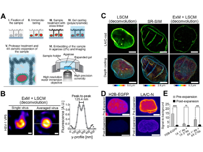
Graphical presentation of pre-ExM sample preparation method and influence of ExM on detection of intranuclear protein complexes and signal fluorescence intensity. Credit: Elina Mäntylä et al.
Researchers from Tampere University as well as The Ohio State University have developed nano-sized force sensors that can measure intracellular forces and mechanical strains on proteins within a cell. The force sensors will allow researchers to observe the forces on a much smaller scale than was previously possible.
Despite being possible to measure forces experienced by individual proteins, analyzing intracellular forces has not been possible until now. With development beginning in 2019, the team of researchers has developed a novel force sensor that can measure these forces on a very small scale. The study, which is published in Molecular Biology of the Cell, outlines the development of the sensor as well as analysis of the intracellular forces.
“The power-sensing part is like a rubber band that changes colour when stretched. This part is attached to the antibodies at both ends of the rubber band, which bind to the cellular target protein under study. The force or elongation of the studied protein can then be detected under a microscope by following the elongation of the rubber band, i.e. the colour it produces,” says Teemu Ihalainen, a Senior Research Fellow at Tampere University
The researchers believe that the new sensor, which is only 20 nanometers, can be used in a variety of cell biology applications. It will allow researchers, for the first time, to be able to visualize the mechanics of an individual cell. To visualize the results of the force sensor, the researchers utilized super-resolution microscopy, specifically expansion microscopy. Initially, the expansion microscopy resulted in a lower-than-desired resolution, but the researchers discovered that by adding more fluorescent labeling the resolution was improved.
“In practise, what we did was to pump more fluorescent molecules to the target proteins as if we were adding reflectors. The simple and easy method greatly improved the resolution and contrast of the image. Noise was also computationally removed from the images, which further increased the image sharpness,” Ihalainen said.
The findings of the study allow future researchers to better understand the basic mechanics and forces that occur within a cell, further research will also lead to better generalizing the technique for use in various applications within cell biology.