Schematic diagram of 3D-SPI technique. Credit: LIU Yifan et al.
Researchers from the University of Science and Technology (USTC) of the Chinese Academy of Sciences (CAS) have developed a method of volumetric imaging of microscopic objects with a near-diffraction-limit 3D optical resolution. The method provides a streamlined approach to imaging with improved spectral range and timing response.
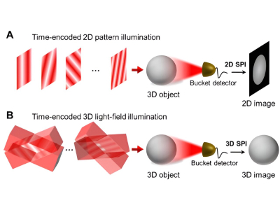
Published in Proceedings of the National Academy of Sciences (PNAS), the novel imaging method called three-dimensional single-pixel imaging (3D-SPI) is based on 3D light-field illumination to enable volumetric imaging. To prove the technique, the team of researchers was able to image single algal cells in vivo.
SPI has recently garnered attention as a 3D imaging method thanks to its improved spectral range and resolution. SPI utilizes single-pixel detectors instead of a detector array of conventional imaging methods, allowing for precise in vivo imaging. 3D-SPI typically relies on time-of-flight to extract depth information, however, this has only been able to obtain millimeter-level imaging at best, far from the microscopic level needed for cellular imaging.
To obtain the microscopic level needed for cellular imaging, the researchers built a 3D-LFI-SPM (or 3D light-field illumination single-pixel microscopy). The prototype was able to achieve an imaging volume of ~390×390×3,800 μm3 with a resolution of 2.7 μm laterally and 37 μm axially. Using the prototype, the researchers were able to achieve label-free 3D imaging of living Haematococcus pluvialis cells.
The new methodology provides a means to conduct highly accurate 3D SPI for biomedical research including monitoring cell morphology and growth in situ.