X-ray magnetic circular dichroism imaging of isolated skyrmions. Credit: Chen Luo et al.
Researchers at Helmholtz-Zentrum Berlin für Materialien und Energie in Berlin have developed a method utilizing scanning transmission X-ray microscopy to distinguish the domain wall type of skyrmions. Determining the domain wall type of the materials allows researchers to further characterize suitable materials for future applications.
Magnetic skyrmions are tiny swirling magnetic excitations that are commonly used in spintronic devices such as data storage devices. Current methods made manipulation of them at room temperature a difficult process to control.
With potential uses in information technology, skyrmions are of particular research interest. Of these, ferrimagnetic rare earth-transition metal (RE-TM) materials are at the forefront of investigation as they exhibit tunable properties including controllable magnetism paired with perpendicular magnetic anisotropy. One class of alloys with particularly strong perpendicular magnetic anisotropy contains a compound of dysprosium (Dy) and cobalt (Co) and has the potential to store information in a more stable way than is currently possible.
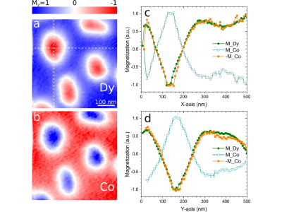
In the study, published in Communications Physics, researchers utilized scanning transmission X-ray microscopy to distinguish the domain wall type of various materials by exploiting the fact that RE materials have stronger linear dichroism than transitional metals. “This allowed us to directly observe isolated ferrimagnetic skyrmions in high density and to accurately determine their domain wall type," said Florin Radu physicist at BESSY II. These findings will allow researchers to reliably distinguish wall types, allowing for the discovery of additional applications of these materials in spintronic devices.