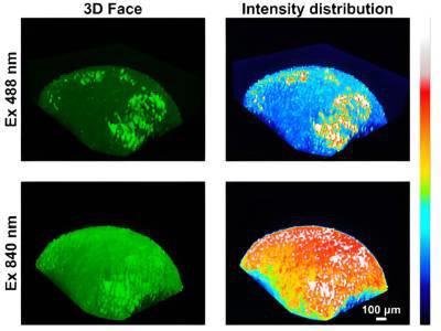
Credit: Jiajie Diao and Yuije Sun.
Two researchers from the University of Cincinnati are pushing the boundaries of imaging by jointly researching and developing new technologies and methods. One of their recent developments, called two-photon imaging, will allow researchers to penetrate deeper to better visualize organelles within human or animal cells.
The research duo at UC, Jiajie Diao and Yujie Sun, have employed a simplistic approach to their research. The two allow the data to take them where it might during a project, avoiding any preconceived notions of what may come of the research. The approach has been successful, resulting in numerous advancements and publications in journals such as Chemical Science and Biosensors and Bioelectronics.
Previous probes developed by the team to focus on lysosomes were successful at providing more detailed information, however, due to the short wavelengths of light that activated them they were not strong enough to penetrate tissue. "We had to use shorter wavelengths to excite the probe, and normally it's visible wavelengths, so their penetration is really limited," said Jiajie Diao, associate professor at the University of Cincinnati. "We could not go deep, so it's good for cellular imaging, but it's not good for tissue or research in living organisms (in vivo)."
The most recent advancements from the team include utilizing two lower-energy photons that can penetrate deeper thanks to longer wavelengths. "Many times, tissue or even in vivo measurement is more important than cellular," said Diao. "This probe will illuminate individual organelles, and it's much better than the commercial two-photon probes currently available."
The team has also developed a probe that can be used to measure individual cell viability. The probe initially stains a cell's mitochondria, however, when that cell becomes damaged the probe will move itself and stain the nucleus. "Eventually the signal on the mitochondria will start getting dim, and the signal on the nucleus will get stronger, so by measuring the color intensity ratio between the mitochondria and the nucleus, we can quantitatively assess the viability of individual cells," said Diao.
The two researchers have been prolific in making advancements and publishing their research since joining forces. "I think the most important thing is always keeping an open mind, communicating frequently and not getting limited," said Diao. "We always say data is data. Many times the biggest fight will be the fight between some preset imagination."