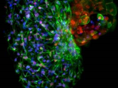
Hepatocyte spheroid, imaged with Lightsheet Z.1. Image cropped. Credit: ZEISS Microscopy via Flickr Commons, CC BY-NC-ND 2.0. Sample courtesy of TNG Weiquan John, Ng Huck Hui, Genome Institute of Singapore. Original Image
Live-cell imaging enables researchers to observe the intricate dynamic changes that are essential to the development and function of organisms, observations that can ultimately lead to a better understanding of human health and disease, and aid in the development of effective therapies for cancer, autoimmune disease, cardiovascular disease, neurological disorders and more. Live cell imaging comes with unique challenges compared with imaging of fixed and cleared biological samples, including the need for high temporal resolution and methods that minimize toxicity to the sample, including phototoxicity.
Two microscopy techniques that have grown popular for live-cell imaging in recent years are light sheet fluorescence microscopy (LSFM) and super resolution microscopy, each offering their own advantages over other techniques like standard wide-field fluorescence microscopy and confocal microscopy. Each of these techniques also involve their own limitations and technological requirements that must be weighed when considering a method for your own live-cell imaging experiments. This article will provide summaries of both approaches to live-cell microscopy, highlight some of their key differences and help bring you a step closer to understanding which method is best suited for your specific application.
Light Sheet vs. Super Resolution
Light sheet fluorescence microscopy (LSFM), also known as selective or single plane illumination microscopy (SPIM), is a microscopy method that utilizes an excitation beam with a cylindrical lens to illuminate a thin section of a sample at a time, rather than flooding the entire sample with light. The LSFM instrument includes a separate excitation objective and detection objective that are oriented perpendicularly to each other, so that the overlapping area between the excitation and detection beam is captured.
Super resolution microscopy encompasses multiple different techniques that allow images to be captured at a higher resolution than typically possible due to limitations defined by the diffraction limit. While there are dozens of methods that can be described as super resolution techniques, this article focuses on three of the most common used in fluorescence microscopy—structured illumination microscopy (SIM), stimulated emission depletion microscopy (STED) and single-molecule localization microscopy (SMLM).
Differences in Phototoxicity
Assessing phototoxicity is essential for developing any microscopy method that requires live samples, as dead cells are the last thing you want to see in your live-cell images. Decreased phototoxicity is one of LSFM’s greatest benefits, as only a thin section across the focal plane is illuminated rather than the entire sample. This allows samples to be imaged for long periods of time to produce comprehensive 3D images and videos without causing serious photodamage.
Super resolution techniques carry a higher risk of phototoxicity than LSFM, as they require higher illumination intensities and a higher number of image acquisitions. SMLM techniques suffer the most in this area and remain challenging to apply to live cell imaging, limiting them to specific applications requiring the exceptional spatial resolution afforded by methods like stochastic optical reconstruction microscopy (STORM) and photo activated localization microscopy (PALM).
SIM is considered to be the least phototoxic of the super resolution methods and is regularly used for live-cell imaging with success.1 Advancements reducing the number of exposures required to generated a reconstructed SIM image, and implementations of new SIM-based techniques that reduce the intensity or area of illumination, have further reduced phototoxicity and enabled longer live-cell SIM experiments to be performed without sample destruction or photobleaching. One example is SIM-total internal reflection fluorescence (TIRF), in which only a few hundred nanometers of the coverslip is illuminated, which reduces photodamage to the sample but also restricts observation to regions close to the coverslip. Another advancement is the development of deconvolution algorithms that can produce super resolution images from SIM data acquired at a lower signal-to-noise ratio, which means a lower illumination intensity can be used.
One difficulty with using STED for live-cell imaging is the use of a high-intensity depletion beam, which carries a high risk of phototoxicity. However, this technique can also be adapted to be more live-cell friendly. For example, using a 775 nm depletion beam has been found to reduce phototoxicity and is compatible with a number of fluorescent proteins and probes.2 Using fast resonant scanning mirrors to increase scanning speed, and using two-photon excitation to limit excitation to the focal volume of the beam, have also been used to reduce phototoxicity in live-cell STED experiments.1
Differences in Spatial Resolution
Super resolution microscopy is what it says on the tin – it provides superior spatial resolution to standard light sheet microscopy, which is constrained by the diffraction limit. It is still possible to achieve cellular-to-subcellular resolution in live samples with LSFM depending on factors such as light sheet thickness, beam shape and the NA of the detection objective, but you will not be able to resolve finer nanoscopic details such as structures within organelles or individual proteins using a standard LSFM. Super resolution techniques, on the other hand, break the diffraction barrier and can provide twice the resolving power of other fluorescence microscopy techniques, or more, depending on the type used.
SIM generally doubles the spatial resolution of conventional wide-field techniques, achieving lateral resolutions down to 100 nm in live samples for linear SIM methods.3 STED and single-molecule techniques like STORM and PALM can both reach lateral resolutions down to 50 nm for live cells, although SMLM techniques can even reach resolutions of 20-30 nm depending on the properties of the fluorophores used.4
A good question to ask yourself when you’re considering using a super resolution system is how high of a resolution you really need in order to achieve the goals of your experiment.5 There are many live-cell imaging experiments that will not require nanoscopic vision in order to observe the structures and processes you want to analyze, in which case light sheet microscopy would be a suitable, or better, choice based on other factors such as speed and cost.
Differences in Temporal Resolution
You will want to maximize your temporal resolution in live-cell imaging experiments when you want to capture the dynamic processes going on inside cells. In this area, LSFM has many advantages, achieving high acquisition speeds and capturing high-resolution 3D images much faster than point-scanning methods. Compared to confocal microscopy, LSFM essentially parallizes data acquisition by capturing all the pixels in a single plane simultaneously, boosting temporal resolution about 100-fold over confocal, with the maximum frame rate determined by the camera being used.6 This allows LSFM to easily capture rapid biological processes such as the beating of a zebrafish heart or the firing of neurons using calcium reporters.
Super resolution techniques require more captures to reconstruct a full image and thus it is difficult to achieve temporal resolutions comparable to LSFM using super resolution techniques without special adaptations. SMLM methods in particular require thousands of captures to produce a super resolution reconstruction and are generally not preferred for imaging live-cell dynamics.2 On the other hand, SIM requires fewer images per time point and can be used effectively for creating time-lapse videos of live-cell processes, with the temporal resolution largely dependent on the setup being used. Traditional SIM setups have been used to produce 3D live-cell videos with frames a few seconds apart, but implementations like SIM-TIRF have improved temporal resolution to 11 Hz or more, with acquisitions as fast as 100 Hz reported using this setup in combination with other optimizations.
While STED is a point-scanning method and requires more time to acquire full images than parallelized methods like LSFM, it does have the advantage of acquiring images in real-time rather than relying on computational post-processing to produce images. Applications of STED with 2 s time resolution have been reported, which is sufficient for observing processes such as organelle dynamics and protein trafficking.2
Differences in Cost
Cost will no doubt be a major factor in considering whether to implement a light sheet or super resolution system. LSFM does require a dedicated system and is a significant investment if you are implementing it for the first time – the cost of computer infrastructure to support large amounts of LSFM data should also be considered. However, LSFM is still much less costly than dedicated super resolution systems. SIM imaging requires a dedicated system, and STED can be performed using either a dedicated system or by upgrading a confocal microscope to perform STED, if such an upgrade is available, which also comes with a significant cost. One exception is SMLM, which can often be performed using an already available fluorescence microscope in conjunction with proper lasers, cameras, reagents and software to perform single-molecule localization. SMLM methods like STORM are also available as complete instrument systems, which increases the cost but could improve performance and ease implementation while freeing up systems for other experiments.
One especially cost-effective approach to implementing LSFM is the OpenSPIM platform, a modular open-source design to build one’s own compact light sheet microscope using off-the-shelf and 3D-printed parts.7 While this platform significantly reduces the cost of implementing light sheet-based live cell imaging, system limitations and the potential challenges of building and configuring such a system should be considered to determine whether this is a worthwhile undertaking for your research.8
References
- Tosheva, K. L.; Yuan, Y.; Matos Pereira, P.; Culley, S.; Henriques, R. Between Life and Death: Strategies to Reduce Phototoxicity in Super-Resolution Microscopy. Journal of Physics D: Applied Physics 2020, 53 (16), 163001. https://doi.org/10.1088/1361-6463/ab6b95.
- Guillaume Jacquemet, Alexandre F. Carisey, Hellyeh Hamidi, Ricardo Henriques, Christophe Leterrier; The cell biologist's guide to super-resolution microscopy. J Cell Sci 1 June 2020; 133 (11): jcs240713. doi: https://doi.org/10.1242/jcs.240713
- Godin, Antoine G.; Lounis, B.; Cognet, L. Super-Resolution Microscopy Approaches for Live Cell Imaging. Biophysical Journal 2014, 107 (8), 1777–1784. https://doi.org/10.1016/j.bpj.2014.08.028.
- Sigal, Y. M.; Zhou, R.; Zhuang, X. Visualizing and Discovering Cellular Structures with Super-Resolution Microscopy. Science 2018, 361 (6405), 880–887. https://doi.org/10.1126/science.aau1044.
- Richardson, D. Introduction to Super-resolution Microscopy. Harvard Center for Biological Imaging Lunch and Learn Lecture Series, 2018. https://www.youtube.com/watch?v=xpAwquiB6AM
- Sébastien Wolf and Georges Debrégeas, CHAPTER 1: Fast Volumetric Imaging Using Light-sheet Microscopy. Principles and Applications, in Optogenetics: Light-driven Actuators and Light-emitting Sensors in Cell Biology, 2018, pp. 1-24 DOI: 10.1039/9781788013284-00001 eISBN: 978-1-78801-328-4
- OpenSPIM website. https://openspim.org/index
- Girstmair, J., Zakrzewski, A., Lapraz, F. et al. Light-sheet microscopy for everyone? Experience of building an OpenSPIM to study flatworm development. BMC Dev Biol 16, 22 (2016). https://doi.org/10.1186/s12861-016-0122-0