by Pedro Echave, Global Leader and NGS Product Expert,, and Mike Brenan, Global Single-cell Product Expert, Revvity
There is truth in the adage that says, “what you put into things is what you get out of them.” In laboratory science and especially in single-cell research, this is certainly true.
Beginning with a high-quality sample will help ensure quality results downstream and thus, more meaningful biological insights. There are inherent challenges, however, in working with single-cell samples. For one, the cell types being studied are especially fragile and more prone to damage or death during collection, storage and sequencing. Fortunately, there are myriad solutions available to aid research teams that encounter these challenges in their single-cell RNA sequencing (scRNAseq) studies. A clearer understanding of what causes low sample viability, how viability affects scRNAseq data and what can be done to prevent low viability in the first place will set research teams on the right track.
The State of Single-cell Research
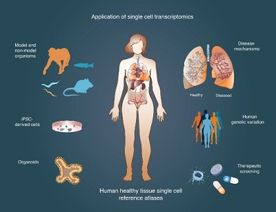
Due to what some have termed the “cellular resolution revolution,” single-cell analysis has evolved from a niche approach to a well-established methodology with broad appeal. The various techniques developed to conduct single-cell studies enable scientists to drill down to human health and disease at the most basic level – uncovering information that informs how certain conditions can be detected and treated. One group making impressive headway in this field is the Human Cell Atlas (HCA), an international consortium founded in 2016. Using high-throughput single-cell technologies, the HCA community is creating what is akin to a “Google maps” of the human body – defining the position, function and characteristics of every cell type in the human body. According to the HCA’s website, the group anticipates its work to impact almost every aspect of biology and medicine – though most recently its contributions to COVID-19 research are notable (e.g., identifying cells susceptible to infection and developing better understanding around how the SARS-CoV-2 virus attacks cells throughout the body).
In recent years, technological improvements have led to scRNAseq emerging as a preferred method to conduct single-cell research across other areas of innate immunology, infectious diseases and clinical research. These improvements have further optimized sensitivity, turnaround time and helped lower the costs associated with sequencing. In fact, even outside of more sophisticated laboratory settings, remote and low-resource lab locations can adopt certain scRNAseq platforms to conduct their research.
Regardless of research area or technique chosen, the inherent challenges of working with fragile, single-cell samples remain. In addition to sample types being more susceptible to damage, scRNAseq data sets are prone to “noise” from ambient RNA, which makes bioinformatics analysis downstream particularly complex. These and other factors place greater importance on preventing low viability samples that in turn will lead to poor results. Understanding the causes of low viability and what can be done to counter or prevent them can help.
Low Sample Viability – Causes and Effects
Nearly all applications of scRNAseq involve these few basic steps: cell isolation, extraction and amplification, sequencing library prep, next generation sequencing (NGS) and data analysis. In all but the last of these processes, sample quality can become comprised due to any number of manipulations – whether they are intentional or not. For this reason, greater emphasis should be placed on the protocols chosen for each step in the scRNAseq workflow.
With cell isolation, for example, certain disassociation protocol – either mechanical or enzymatic – can be especially harsh for fragile cell types. Among the more challenging cell types to work with are granulocytes – the focus of studies in innate immunology and the most common type of white blood cell (also inclusive of neutrophils, eosinophils and basophils). These sensitive cells produced in the bone marrow live just for a few days in the human body and are especially difficult to recover with certain microfluidic-based platforms.
Sample storage is another common cause of low viability. Cryopreservation is one of the more commonly used techniques to store cells for transportation and evaluation at a later date. In essence, this practice aims to “lock in” cellular structures and expression at very lower temperatures (-80 °C or lower). Even when working effectively, cryopreservation exposes samples to harsh temperatures that can lead to harm or premature cell death. This is especially true if samples are stored in this state for weeks or months at a time.
The consequences of poor sample quality are not poor results alone. Research teams will invest the same amount of time and money to sequence a low viability sample as they would a high viability sample – just with less meaningful results.
How to Assess Sample Viability
Recognizing the importance of quality data in to get quality data out, there is a growing market for solutions to assess sample viability before it can become an issue.
To date, many researchers have chosen to use a hemocytometer to perform manual cell counting. This method involves adding a solution of trypan blue to a sample before examining it more closely under a microscope. Healthy, live cells will be unaffected by the trypan blue, whereas the stain will penetrate the broken membranes of dead cells and allow users to determine overall viability. This method typically involves the least amount of upfront capital investment to perform and is recognized as being fairly robust. Still, as a manual process, it comes with the possibility of human/user error. Seasoned single-cell researchers and newcomers alike may find it challenging to evaluate samples, especially knowing that in some cases, trypan blue can overestimate viability.
As a result, automated cell counters have grown in popularity among single-cell researchers. With some systems available as compact, benchtop devices, automated cell counters offer greater efficiency and reproducibility for teams looking to assess the viability of multiple samples. This has added value for researchers pursuing multi-point or longitudinal studies.
Options to Optimize (or Altogether Prevent) Low Viability Samples
If a sample is found to have low viability, researchers are not out of options. There are a number of cell enrichment kits on the market that aim to provide more starting material from what might otherwise be disregarded as a low viability sample. Underlying technologies in these solutions vary, but many focus on cell density and use magnetics to separate live from dead cells. Researchers should keep in mind that despite being classified as “enrichment” solutions, these can work the opposite way too. These kits introduce an added manipulation to a sample that can negatively impact viability.
As an alternative to cell enrichment, researchers can instead choose to focus on optimizing their scRNAseq workflow upstream. Selecting protocols that allow for faster cell isolation, extraction and amplification are ideal, as is optimizing the physical setup of the laboratory to expedite these processes. When possible, using scRNAseq platforms that avoid common manipulation points altogether could be considered.
When enrichment and upstream improvements aren’t possible, scRNAseq studies can still proceed with low viability samples. In these instances, research teams will choose to move forward with low viability samples knowing that the data output will not be the best possible. In these unideal cases, more careful analysis with the support of bioinformatics experts can still yield meaningful findings to inform future research.
Implications
In the coming years, we can expect single-cell analysis to continue growing in popularity – propelled by new technologies and automated workflows. The acceleration and simplification of translational, pre-clinical and clinical research will in turn yield new insights in oncology, nephrology, neurology and other areas of medicine, ultimately benefiting the clinical community and improving human health.