A corneal transplant. Credit: VisionGift
by Kristin Mathes, MS, MA, VP of Quality Systems, VisionGift
A group of criminals recently broke into a bank by cutting the power to the facility to avoid tripping any alarms. They went for the goods, only to discover that it wasn’t a financial bank. They had broken into an eye tissue bank.
Although the thieves were shocked to discover what was actually housed at the facility, the real issue at the tissue bank was the loss of power to the storage chambers. With the temperature in one of the tissue storage freezers rising out of its specified range, the ocular tissues were at risk of damage.
Fortunately, an immediate notification through a continuous monitoring system alerted VisionGift, a nonprofit dedicated to advancing sight for all humankind through eye donation, transplantation, and research, that something was wrong and to act quickly to protect the tissue destined to give the gift of sight through corneal transplantation.
In tissue banks and other critical environments, advanced monitoring systems constantly assess and analyze crucial parameters like temperature, humidity, and other vital factors in real time to help prevent potential disasters and safeguard mission-critical work.
Tissue banks and innovations in allograft technology
Tissue banks serve as repositories of a diverse range of tissues, each with the potential to contribute to groundbreaking research, life-saving transplants, and medical advancements.
Through the preservation and utilization of donated tissues, these specialized facilities stand as beacons of progress, driving advancements that have a lasting and positive influence on the health and well-being of communities worldwide.
Ocular tissue banks play an invaluable role in restoring sight and transforming lives through innovations in corneal transplantation and allograft technology. Each year, nearly 50,000 corneal transplants occur in the United States and roughly 80,000 worldwide. Corneal transplants—aka keratoplasty procedures—are life-changing for patients with impairing conditions like keratoconus or infections.
By reshaping or replacing damaged corneas, surgeons can correct visual problems that glasses and contacts cannot. With a success rate exceeding 95%, corneal transplants allow many blind or visually impaired individuals to regain independence and mobility while improving their quality of life.
There are three common types of corneal transplants:
- Endothelial keratoplasty replaces the innermost layer of the cornea called the endothelium. This layer can be damaged by conditions like Fuchs’ dystrophy that cause swelling and cloudiness. Replacing just the endothelium preserves structural integrity better than a full-thickness corneal transplant.
- Penetrating keratoplasty involves a full-thickness transplant of all the corneal layers. This kind of transplant is performed for disorders affecting the entire cornea, like keratoconus or infections.
- Anterior lamellar keratoplasty replaces the outermost corneal layers, including the epithelium and stroma. This approach spares the endothelium and is useful for surface diseases like corneal scarring.
These procedures can treat disorders like keratoconus, where the cornea thins and bulges into a cone shape that progressively impairs vision. As the condition advances, the cornea becomes too thin and irregular for vision-correcting glasses or contacts to work, resulting in blindness. Corneal transplantation reshapes the cornea to restore more normal vision.
New allograft technologies are advancing treatments, as well. KeraNatural, developed by VisionGift, uses an irradiated corneal graft to structurally reshape a keratoconic cornea. The graft is shaped and inserted into the recipient cornea through a tunnel created with a femtosecond laser. Developing specialized allografts allows tissues to be used creatively and can increase the number of lives one donor can impact.
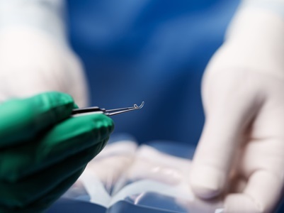
In VisionGift laboratories, donated corneal tissue is cut into a specific shape to form a graft and then sent for irradiation. Once in the hands of the surgeon, the graft is inserted into a laser-cut tunnel in the patient's cornea. The graft alters the shape of the cornea by flattening the coned or bulging area and normalizing the curvature for improved vision. They may also use laser surgery to fine-tune the eye's surface with the graft. It’s truly some of the most exciting eye surgery in the ophthalmologic world right now.
Innovations that expand the use of donated tissues also honor the selflessness of donors and their families. By maximizing each donation, vision restoration aids more recipients. However, realizing these innovations requires strict compliance with standards to progress responsibly.
Compliance with rigorous standards
Eye banks must maintain accreditation with the Eye Bank Association of America and comply with regulations across multiple jurisdictions, including the U.S., Canada, the European Union and the United Kingdom.
Compliance with rigorous standards in eye banks is crucial for ensuring the safety, efficacy, and quality of ocular tissues while advancing medical science, and improving lives. Adherence to established standards guarantees the quality and safety of corneal tissues used in transplants, increasing the success rate and restoring vision.
When ocular tissue is donated, it goes to the laboratory to be evaluated for either short- or long-term storage. Timely evaluation and appropriate designation help ensure the optimal use of donated tissues for various medical purposes. The designation of tissue for short- or long-term storage is largely determined by its endothelial integrity.
Short-term storage tissue, referred to as “fresh” tissue, is not irradiated and has living cells necessary for 99% of surgeries that use this type of tissue. Surgeries involving endothelial keratoplasty and penetrating keratoplasty rely on healthy endothelium. Tissues lacking functioning endothelial cells are instead evaluated for long-term storage.
Tissue designated for long-term storage is sterilized by electronic-beam irradiation. Frozen corneal tissue is kept in media containing recombinant human serum albumin for long-term storage. After irradiation, the tissue can be safely stored at room temperature for up to two years, enabling flexibility in scheduling less time-sensitive procedures.
The clock starts ticking as soon as the tissue has been recovered. Ophthalmic surgeons are extremely particular about the length of time between the date of death and the date of surgery for transplants using fresh tissue, typically only using tissue with no more than several days in media. This requires quick action, especially if the tissue goes overseas. But, if the donation is acceptable for long-term use, we can store it for up to a year in -80° C freezers before irradiation, giving us a larger window to honor the gifts from a donor and ensure we are good stewards.
Strict timelines must be followed when utilizing fresh tissues before cell degradation because even minor delays can render transplants unviable. Consequently, continuous monitoring is essential for maintaining compliance in these sensitive tissue bank environments and avoiding disruptions—such as a loss of power.
The value of continuous monitoring
In the VisionGift case, when the would-be thieves cut power, HVAC systems and the network shut down. To mitigate any damage while waiting for conditions to return to normal, the team kept all storage chambers closed.
Receiving remote email, text and phone notifications from the Vaisala viewLinc Continuous Monitoring System, the team realized that one of the freezers was not returning to its correct temperature. Unchecked, the temperature would quickly climb above the safe range, destroying the precious corneal tissues within. Consequently, it was essential to get to the facility quickly to intervene.
Upon arrival, the team checked the data logged on the monitor’s sensors during the outage, confirming enough time to move the affected tissues into a functioning freezer before any impairment occurred. But if VisionGift relied on manual periodic monitoring, far more tissues could have been damaged before a problem was discovered.
Continuous monitoring sensors track conditions 24/7 and log high-resolution data rather than periodic manual readings. Intelligent algorithms distinguish environmental fluctuations from temporary spikes, with tight alarm thresholds warning of minor deviations before they become critical. Plus, remote access to data allows rapid response to alerts from any location, and backup power keeps systems running even when local power fails. Finally, carefully designed reporting provides meaningful oversight for compliance and tissue viability assessments.
The continuous data also helped confirm the viability of the tissues after the incident. Accelerated warming can make tissues unusable even if temperatures don't exceed thresholds for very long. Members of the VisionGift team consulted with an infectious disease expert, providing the time-stamped records to assess whether the temperature fluctuations caused invisible damage. Thankfully, the tissues were confirmed as safe.
Driving transplantation innovation
Advanced monitoring systems are critical in facilities far beyond tissue banks. Any environment maintaining tight controls over sensitive materials can benefit, from eye banks to pharmaceutical and biotech labs, data centers, art galleries, museums and beyond.
Data empowers informed decision-making while uptime assurances keep vital operations running smoothly. In the end, environmental monitoring helps organizations focused on advancing science, improving lives, and serving humanity. The story of VisionGift is just one example of the importance of continuous monitoring, but similar stories unfold nearly every day in facilities across the globe. Wherever materials vital to human health are stored or produced, environmental monitoring stands guard.
So, while the alarms that alerted VisionGift to trouble may have felt disastrous in the moment, they prove that sophisticated technologies vigilantly watch over one of our most cherished gifts—that of sight.