Featured Article
Biological microscopy is benefitting from advances in technologies, software, and equipment at a rate that is allowing researchers to see even deeper, and more clearly, into the dynamics of living cells—the subtlest details and processes that define life. Today scientists are using powerful super-resolution microscopy techniques to image living samples well beyond the diffraction limit of optical microscopy, allowing fine mapping of cellular structures and specimens that are incompatible with electron or atomic force microscopy.
As super-resolution technologies evolve, they become more user-friendly across a wide range of applications. This has led to a group of techniques aptly named functional super-resolution microscopy. Some functional super-resolution techniques have even undertaken speed and phototoxicity concerns, making them appropriate for live cell imaging experiments.
For these reasons, functional super-resolution techniques have largely become the biological imaging method of choice when super-resolution is needed (see Figures 1 and 2). There are two major categories in which the various super-resolution techniques fall: deterministic and stochastic.
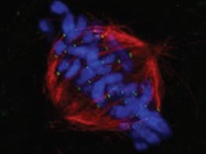
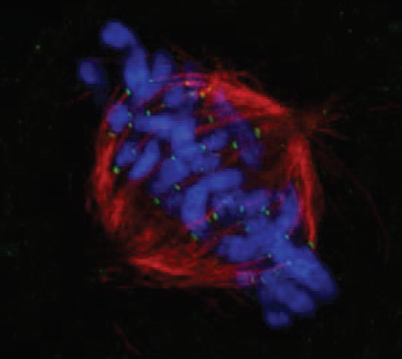 Figure 1 – HeLa DAPI a-tubulin Hec1. Credit: Masanori Ikeda and Kozo Tanaka, Department of Molecular Oncology, Institute of Development, Aging and Cancer, Tohoku University.
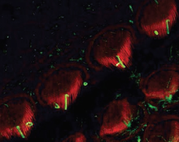 Figure 2 –
 Stereocilia Kinocilia
. Credit: Hatsuho Kanoh and Toru Kamitani, Graduate School of Frontier Biosciences and Graduate School of Medicine, Osaka University.Functional super-resolution
The principal process of functional super-resolution is localizing the coordinates of many diffraction-limited emitters (e.g., single fluorescent molecules), fitting their point-spread functions (PSF), and reconstructing the localizations to create a final image of the specimen. Stochastic super-resolution techniques achieve this through a series of fluorophore activations and deactivations that localize the coordinates of single molecules. This process is repeated for thousands of frames as images are acquired point-by-point. These methods include photoactivated localization microscopy (PALM) and stochastic optical reconstruction microscopy (STORM).
Deterministic super-resolution methods like stimulated emission depletion (STED) and ground state depletion (GSD) exploit optical responses to excitation light to enhance resolution. These techniques can resolve structures down to 10 nanometers, providing astonishing accuracy under the right imaging conditions. However, because they often require the use of complex hardware, special fluorophores, and imaging buffers, these techniques are out of reach for many traditional imaging experiments.
Imaging the invisible
Structured illumination microscopy (SIM) separates and reconstructs the Fourier transform of an image by shifting several frames in an image by some phase, utilizing image information disregarded in widefield imaging. Saturated structured illumination microscopy (SSIM) is a deterministic super-resolution technique that builds upon this shifted-phase, multiple-exposure principle through a sinusoidal wave pattern, taking advantage of fluorophore emission rate dependence on laser intensity.
Additional benefits of super-resolution microscopy
Other advantages of modern super-resolution microscopy include:
High-speed data processing algorithms: These enable super-resolution images to be viewed in a live display window to observe live cells, various cell structures, and other molecules. Advances in GPU processing remove the barriers of temporal resolution, making real-time super-resolution possible with confocal SIM for imaging rapid intracellular dynamics.
Observation at depth: Super-resolution microscopy allows the study of subcellular architecture and dynamics at the nano scale. Researchers can clearly observe not only the surface of the sample, but also up to 100 microns deep within the sample.
Three-dimensional imaging: Researchers can obtain detailed 3-D super-resolution image data during time-lapse imaging due to higher temporal resolutions.
Ease of use: Super-resolution microscopy combines intrinsic optic sectioning with fast data acquisition and dual-color super-resolution to provide quality images in a timely fashion for further actions.
Potential super-resolution challenges
Despite its enormous power and potential, there are some inherent challenges associated with super-resolution microscopy, most of which increase as resolution becomes higher. Potential drawbacks include:
Point of diminishing returns: As super-resolution systems image smaller and smaller details, the population of responding fluorochromes decreases, requiring the development of new fluorochromes with higher quantum yields. The quality of super-resolution images can also vary tremendously; images captured using the same resolution can appear dramatically different in detail and appearance.
Sample reliability: Some live samples are more adversely affected by super-resolution imaging than others because of high excitation intensity or extended exposure times. Stress placed on the specimen can also have an impact on data reliability, which can potentially outweigh the benefits of higher resolution.
Optical aberration: Differences in refractive index have significant implications on the optical properties of light. Proper use of immersion oils designed to properly match the refractive indices of both live and fixed samples is critical to achieving the highest possible resolution.
Considerations and possibilities
As users weigh pros and cons, they should review the specific needs of their research, consider the validity of all solutions, and investigate all super-resolution techniques and other microscopy methods, including widefield and confocal imaging. The best choice is the method that produces the most usable fluorescence emission while providing all of the necessary data. With confocal imaging, lowering the background intensity can improve fluorochrome visualization and enhance image analysis. In some cases, this enhanced contrast is enough to provide sufficient resolution. In other instances, super-resolution is required.
As technology advances and software becomes more sophisticated, optical and computational super-resolution techniques are coming together in commercial imaging instruments to offer flexible, multimodal systems capable of producing images beyond the diffraction limit. Some imaging systems are available that allow the use of confocal and super-resolution modes within the same system. These systems allow the operator to select the mode that will provide the best results for a given application and provide the advantage of a common stage. Using the same slide, the researcher can switch back and forth between confocal and super-resolution modes, producing two data sets for comparison. This added benefit allows multiple sources of information that can help confirm findings and lead to better certainty without artifacts.
While there are many functional super-resolution techniques that offer various advantages and limitations, the primary benefit of super-resolution is enhanced resolving power. With today’s commercial microscopes offering a variety of user-friendly technologies, super-resolution is now within the reach of lab staff without extensive optical backgrounds. With this new level of imaging support, experiments that once seemed out of the question are now a possibility. Fewer experimental sacrifices need to be made, and existing fluorophore labeling protocols can be used to integrate super-resolution technologies into workflows without disruption.
Lauren Alvarenga is associate product manager, Olympus Corporation of the Americas, Scientific Solutions Group, 48 Woerd Ave., Waltham, MA 02453, U.S.A.; tel.: 781-419-3630; e-mail: [email protected]; www.olympus-lifescience.com