By Brendon Sharp, Science Writer
Fourier-transform infrared (FTIR) spectroscopy is commonly used to analyze organic compounds synthesized by chemists, but reflectance-based FTIR methods can also be a powerful technique for analyzing thin films on reflective substrates. This lesser-used technique can provide useful information for chemists and materials scientists working with thin films on reflective substrates, such as photoresists used in semiconductor fabrication.1 When using FTIR to analyze thin films, there are some additional considerations that conventional transmission/absorption FTIR spectroscopy does not involve. Here are some useful “do’s and don’ts” to maximize the quality of your thin-film FTIR spectra.
DO: Use a highly reflective substrate
Sufficient light must be reflected from the substrate in order to capture a high-quality spectrum. Typically, this requires a metallic substrate such as a gold-coated wafer, but it is also possible to use silicon wafers. However, many more scans (see #3) may need to be collected compared with a more highly reflective substrate such as gold due to a weaker signal.
DON’T: Use thick films
Because the reflectance of IR light is low using this technique, excessively thick films will absorb too much light, preventing it from being reflected back to the spectrometer. Thin films (< 1 µm thick) are generally necessary to ensure that sufficient light is reflected back into the objective.
DO: Collect many scans
Very little light is reflected from the substrate, which generates a weak signal. Therefore, a sufficient number of scans is necessary to collect a useful spectrum. The optimal number of scans will depend on various factors, including the thickness of the film and the reflectivity of the substrate. More reflective substrates and thinner films will generally require fewer scans (i.e., a shorter analysis time) to collect usable spectra. Conventional transmission spectroscopy usually requires only 32 scans per spectra, but with reflectance spectroscopy, it is often necessary to collect 100 or more scans per spectrum in order to adequately resolve peaks of interest.2 This means that each spectrum can require several minutes to obtain, which is much longer than traditional transmission spectra using KBr pellets.
DON’T: Analyze IR-inactive films
As with traditional FTIR transmission spectroscopy, the reflectance technique only works when the analyte is composed of compounds that are IR-active. Therefore, IR-inactive species cannot be analyzed using this technique unless they undergo a chemical change to render them IR-active.
DO: Be mindful of the relationship between penetration depth and wavelength
There is an inverse relationship between wavenumber and penetration depth, in which the penetration depth of the infrared beam increases with the wavelength.3 This gives rise to notable differences between reflectance spectra and transmission spectra. For example, the well-known strong and broad hydroxyl (-OH) peak appears as a much weaker (but still broad) signal in reflectance spectra. Many software packages have a correction algorithm that accounts for this variation and can scale the spectra as necessary.
DON’T: Analyze the edges of the film
Because spin-coated films are often thicker at the edges, spectra should be obtained away from the edges and closer to the center.
DO: Ensure that any solvents have evaporated before analysis
If an organic thin film is deposited using spin coating, the film must be completely dry before FTIR analysis, as most organic solvents will display absorption peaks in the FTIR spectra. Solvents with a higher boiling point will naturally take longer to evaporate from thin films. A bake or a vacuum oven treatment may be necessary to remove any solvents before analyzing thin films using FTIR.
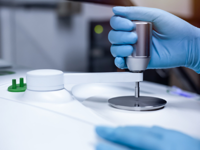
DON’T: Handle thin film samples with hands
Thin films are typically handled in laboratories that use various solvents and chemicals, which can be deposited by handling samples with hands—gloves or no gloves! Due to the thinness of films, any substance that is deposited on the surface may lead to the appearance of unknown bands in the FTIR spectra. Even worse, it’s not always possible to remove these substances from the surface of the film without damaging the sample. Therefore, clean forceps (tweezers) should always be used to handle thin film samples at their edge to minimize contamination and damage risks.
DO: Analyze multiple locations
If you are interested in tracking changes in the chemical composition of a thin film after different processing steps, it’s important that you analyze multiple locations. In an ideal world, thin films would be homogeneous, but they are typically multi-component formulations, which can lead to aggregation of formulation components, such as photoacid generators and other additives. This may ultimately lead to slightly different chemical compositions in different regions. Obtaining spectra from multiple locations can help reveal these differences.
DON’T: Forget to collect a background spectrum
As with any spectroscopic technique, a background spectrum must be collected, generally using an uncoated, bare substrate such as a silicon wafer. For this technique, it’s important to collect a background signal using the same parameters as the measured spectra. Because spectra are collected in the ambient atmosphere using this technique, a background spectrum may need to be collected between measurements due to changes in ambient humidity or CO2 levels due to the presence of the operator.
DO: Use smooth, continuous films
Although not as sensitive to roughness as similar thin-film analysis techniques such as UV-Vis and ellipsometry, rough surfaces may still diffusely reflect light and prevent some of the IR light from penetrating the film. Often, air bubbles and particles will appear on the surface of a thin film after spin coating. If possible, avoid measuring FTIR spectra near these areas.
About the author
Brandon Sharp, Ph.D., is a freelance technical content writer with experience designing photoresists and other organic materials for advanced lithography applications.
References
- Rosenthal, P. A.; Xu, J.; Charpenay, S.; Cosgrove, J. E.; Ravindra, N. M. Infrared Analysis of Advanced Thin Film Materials. JOM-e 2000, 52 (10).
- Thin Film Sample Measurement Methods and Precautions. https://www.shimadzu.com/an/service-support/technical-support/ftir/tips_and_tricks/thinfilm.html (accessed 2023-07-12).
- Q: How deep does the infrared light penetrate at the position of contact between the prism and sample during ATR measurements? https://www.shimadzu.com/an/service-support/faq/ftir/2/index.html (accessed 2023-07-12).