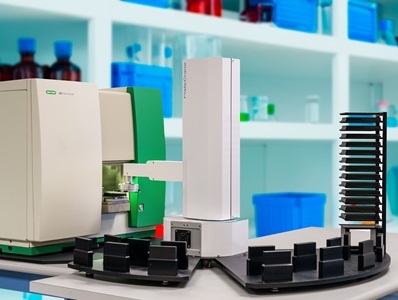
Custom flow cytometry workcell featuring the ZE5 Cell Analyzer with the PlateCrane EX Robotic Arm Microplate Handler and SoftLinx Automation Software
by Richard Cuthbert, PhD, Flow and Antibody Specialist, Bio-Rad Laboratories
As cancer research becomes increasingly focused on understanding the interactions between malignant cells and the immune system, the ability to perform rapid, multiparameter analysis at the single-cell level has made flow cytometry an important component of the oncology research toolkit. Flow cytometry can be used to characterize tumor cells, profile immune populations, evaluate therapeutic response, and support drug discovery programs.
Recent studies specifically highlight how flow cytometry is contributing to the development of novel therapeutic approaches, including metal-based immunotherapies and targeted treatments for hematological malignancies. Advances in instrumentation, assay development, and workflow automation are also enabling researchers to generate larger datasets while maintaining the sensitivity and specificity required for complex oncology applications1.
Exploring the therapeutic potential of metal ions
Metal-containing drugs have played an important role in cancer treatment for decades. Platinum-based chemotherapies remain among the most widely used anti-cancer agents, but researchers are increasingly interested in the broader relationship between metal ions and immune regulation2,3.
This emerging field of metalloimmunology focuses on understanding how metal ions influence immune pathways and how those interactions might be exploited therapeutically. Recent work has shown that metal ions can serve both structural and biological functions, creating opportunities for novel immunotherapeutic approaches that combine drug delivery and immune activation within a single platform3,4. One area attracting considerable attention is the cyclic GMP-AMP synthase-stimulator of interferon genes (cGAS-STING) pathway. This innate immune signaling pathway plays a key role in host defense and anti-tumor immunity by promoting the production of type I interferons and other immune mediators. Activation of STING has therefore emerged as an attractive strategy for cancer immunotherapy5.
Researchers in the laboratory of James Moon at the University of Michigan have investigated how metal ions can enhance STING-mediated immune responses. Their work builds on earlier findings showing that manganese ions can amplify cGAS-STING signaling while simultaneously serving as components of nanoparticle delivery systems. More recently, the group developed a self-assembled coordination nanoparticle known as a zinc-manganese-cyclic dinucleotide (CDN) Particle (ZMCP), which combines metal ions with CDN STING agonists to enhance therapeutic delivery and immune activation5. Flow cytometry played a central role throughout this research. The technique was used extensively for immunophenotyping applications, enabling detailed analysis of immune cell populations in peripheral blood mononuclear cells, spleens, and tumor samples. By monitoring changes in immune-cell subsets and activation markers following treatment, researchers were able to gain insight into the biological mechanisms responsible for therapeutic efficacy5.
In this case, flow cytometry was indispensable for tracking dynamic immune responses and identifying treatment-associated changes within complex immune populations. Such information is critical when developing immunotherapies because researchers must understand not only whether a therapy works, but also how it reshapes the immune landscape to generate an anti-tumor response5. The study showcases how flow cytometry contributes to modern cancer research beyond simple endpoint measurements. By enabling comprehensive immune profiling, the technology provides mechanistic insight that can guide the optimization and translation of experimental therapies.
High-throughput functional assays for targeted therapies
Flow cytometry is equally valuable in the development and evaluation of targeted cancer therapies. Modern drug discovery programs frequently require researchers to analyze large numbers of samples while maintaining sufficient resolution to detect subtle biological effects. Functional assays based on flow cytometry are particularly well suited to these requirements because they combine quantitative cellular analysis with high-throughput sample processing6.
When it comes to the treatment of B-cell malignancies, for example, anti-CD20 monoclonal antibodies such as rituximab have established selective B-cell depletion as an effective therapeutic strategy7. As new biosimilars and next-generation therapies are developed, researchers require rapid and reliable methods to evaluate both potency and specificity.
A recent study described the development of a rapid B-cell-killing assay using peripheral blood mononuclear cells. Mixed populations of immune cells were exposed to a CD20-specific monoclonal antibody and subsequently analyzed by flow cytometry. Using multiple cell-surface markers, researchers were able to identify B cells, T cells, natural killer cells, and monocytes within the same experiment8. The assay demonstrated a significant reduction in B-cell populations following anti-CD20 treatment while leaving other immune-cell populations largely unaffected. Because multiple populations were analyzed simultaneously, researchers could assess both therapeutic effectiveness and target specificity from a single dataset8.
This multiparameter capability is one of the major advantages of flow cytometry, with high-parameter instruments, such as the ZE5 Cell Analyzer, detecting dozens of colors/markers simultaneously, conserving precious samples and saving time. Specifically, instead of performing separate experiments to evaluate different cellular responses, investigators can generate multiple biologically relevant readouts from the same sample. With the speed of analysis being equally important, more than 80 samples can be processed in approximately 10 minutes while collecting over 100,000 cellular events per well. The combination of high event counts and rapid processing provides the statistical confidence needed for screening studies while supporting the throughput requirements of modern drug discovery programs8.
Supporting discoveries across oncology research
The contribution of flow cytometry extends well beyond therapeutic screening and immunotherapy development. The technology continues to play a central role in studies investigating cancer biology, metastasis, biomarker discovery, and treatment response across a wide range of tumor types.
For example, in acute myeloid leukemia, researchers recently used flow cytometry to evaluate a novel proteolysis-targeting chimera (PROTAC) designed to degrade the RNA demethylase FTO. Flow cytometry-based differentiation and apoptosis assays demonstrated that the compound promoted myeloid differentiation and induced significant levels of apoptosis in leukemia cells, supporting its potential as an anti-leukemic therapeutic candidate9.
Flow cytometry has also contributed to advances in breast cancer research. Investigators studying metastatic dormancy used the technique to characterize immune cell populations within synthetic metastatic niches and to track neutrophil subsets associated with suppression of metastatic growth. These experiments provided important evidence that specific immune-cell populations can influence whether disseminated tumor cells remain dormant or progress to form secondary tumors10.
In prostate cancer research, flow cytometry was used to investigate the role of low-molecular-weight tyrosine phosphatase in tumor growth and oxidative stress resistance. Cell-cycle analysis revealed changes associated with impaired DNA repair and altered cellular proliferation, helping researchers better understand the biological consequences of targeting this pathway therapeutically11.
These studies highlight the versatility of flow cytometry across diverse oncology applications. Whether measuring apoptosis, tracking immune-cell migration, assessing cell-cycle progression, or performing detailed immunophenotyping, the technology provides researchers with a powerful means of investigating cancer at single-cell resolution.
Last thoughts
Cancer research increasingly depends on technologies capable of generating detailed biological information from complex cellular systems. Flow cytometry continues to meet this need through its unique combination of speed, sensitivity, and multiparameter analysis.
From supporting the development of metal-based immunotherapies to enabling high-throughput evaluation of targeted cancer treatments, flow cytometry is helping researchers answer important biological questions while accelerating therapeutic discovery. As oncology research continues to advance toward more sophisticated immunotherapies, precision medicines, and cell-based treatments, flow cytometry is likely to remain an indispensable tool for understanding disease biology and translating new discoveries into clinical applications.