Optical thermometry can offer useful two-dimensional temperature information for biological research or clinical diagnostics, without the need to make contact with a sample, specimen or patient. Photoluminescence lifetime imaging (PLI) is one technique that can be used to create a 2D temperature map by measuring the luminescence of nanoparticles doped with rare-earth ions. While PLI offers much higher spatial resolution than a typical thermal camera, one of its major limitations is the long amount of time required to image luminescence point-by-point; this technique also requires that the sample remain stationary to achieve the best resolution. However, a new technique developed by researchers at the Institut national de la recherche scientifique (INRS) allows for wide-field, real-time photoluminescence lifetime imaging in just one exposure, opening up new possibilities for diagnostic optical thermometry.
The new technique, called single-shot photoluminescence lifetime imaging thermometry (SPLIT), uses a novel ultrahigh-speed camera system to capture how quickly the luminescence of the lanthanide-doped upconverting nanoparticles (UCNPs) decays in every spatial point at once. Unlike a normal household camera, which captures one image per exposure, or PLI, which utilizes point-scanning techniques, SPLIT captures all the images of a dynamic event into a single snapshot, said lead author Xianglei Liu. This allows the lifetime of the UCNPs to be captured in a moving sample, and because the UCNPs act as “nanothermometers” that undergo luminescence changes in response to temperature, the SPLIT images can provide a detailed 2D temperature map of a biological sample at single-cell resolution. This research was published in Nature Communications.
“Compared to existing thermometry techniques, SPLIT is faster and has higher resolution. This allows a more accurate temperature sensing with both an advanced and economical solution,” said Jinyang Liang, one of the paper’s corresponding authors.
The researchers believe the SPLIT imaging method could be used in diagnostic applications to detect conditions such as skin cancer. Melanomas, especially micro-melanomas, can be difficult to detect, even when invasive techniques such as biopsies are used. SPLIT could be used as a noninvasive method to locate cancerous cells on the skin, which have a higher temperature than normal tissue, the researchers said. Additionally, SPLIT could be used during photothermal cancer therapy to monitor and tune the light dose to target cancer cells without doing unneeded damage to surrounding tissue.
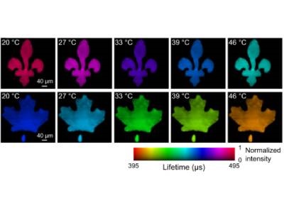
Photo: Lifetime images of green (top row) and red (bottom row) upconversion emission bands under different temperatures captured by SPLIT. Credit: Jinyang Liang