The activities of individual biomolecules such as nucleic acids, enzymes and antibodies can be difficult to observe and track in real time, leaving some questions about their binding behavior and kinetics unanswered. One example of this is the difficulty of differentiating between specific and non-specific binding events in diagnostic analyses of mixed samples like serum. A new microscopy technique developed by researchers at Arizona State University can allow for more precise monitoring of binding activities while simultaneously tracking a total of 100 molecules in 3D.
Surface plasmon resonance microscopy (SPRM) is based on imaging and measuring the disturbance of a surface plasmon by molecular binding activities. A receptor molecule, such as a segment of DNA or an antibody, is affixed to a gold surface, and a binding molecule is then introduced to allow the interactions to occur. When the binding molecules interact with the fixed receptor molecules, the reactions disturb the surface plasmon of the gold film, which is registered as a change in light intensity that is captured by a 400 fps camera. This method allows binding activities between up to 100 molecules to be measured and tracked simultaneously, with sub-nanometer axial precision and single-digit nanometer lateral precision.
One activity the researchers tracked using SPRM is the unwinding of double-stranded DNA by helicase. With the short DNA segment, about 48 base pairs long, fixed to the gold film, the researchers could monitor both unwinding rate and the rotation angle of the helicase molecule as it went to work. The method was also used to observe the binding of the heart disease biomarker troponin T to its corresponding antibody, and could not only detect the antibody-antigen binding interaction but also distinguish between specific and non-specific binding events. This research was published in ACS Sensors.
“Taking advantage of the extremely high sensitivity in the axial direction, SPR can track the particle’s axial motions with sub-nanometer precision, which is much more precise than regular microscopy imaging,” said corresponding author Shaopeng Wang. “We demonstrated that this feature can be used to study the details of molecular binding events at the single molecule-level and also for processing multiple signals at once.”
The ability to capture data on 100 molecules simultaneously using this technique allowed for detailed statistical analysis of binding events and kinetics that would not ordinarily be possible using conventional methods. This technology could lead to improved diagnostic assays and biosensing devices in the future.
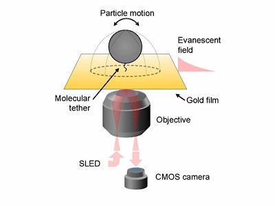
Photo: The graphic shows the basic experimental setup for Surface Plasmon Resonance Microscopy (SPRM). When a particle of interest binds with a receptor, tethered to a thin gold film, the event disturbs a surface plasmon wave, which is registered as a change in light intensity. SLED (for super-luminescent emitting diode) is the light source, which illuminates the sample and induces a surface plasmon wave that ripples across the gold surface. A high speed camera captures these rapid changes, imaging the binding dynamics. Credit: Adapted with permission from Three-Dimensional Tracking of Tethered Particles for Probing Nanometer-Scale Single-Molecule Dynamics Using a Plasmonic Microscope Copyright 2021 American Chemical Society.