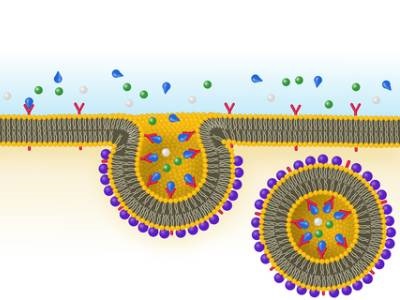
It is well known that cells take in substances such as proteins, hormones, and even pathogens, by forming vesicles on the cell membrane through processes including clathrin-mediated endocytosis, also called receptor-mediated endocytosis (RME). However, the fine details of how the clathrin scaffolds that coat the budding vesicles behave throughout the RME process have been the subject of uncertainty and even controversy for decades. Researchers at Ohio State University now say they have revealed just how the vesicles that cells use to “eat” form, with the help of their clathrin coats, using real-time super-resolution fluorescence imaging of live cells.
Using the high-resolution imaging technology to record video of cell processes, the researchers demonstrated that the formation of curvature by individual clathrin pits could be observed in both cultured cells and in the tissues of developing organisms. Through these observations found that the clathrin coats start causing the membrane to curve beginning in the early stages of their formation, which goes against previous hypotheses that the structure began as a flat lattice and needed to undergo an energy-intensive reorganization process before transitioning from flat to curved.
“Simply put, in contrast to previous studies, we made high-resolution movies of cells instead of taking snapshots,” said lead author Comert Kural. “Our experiments revealed that protein scaffolds start deforming the underlying membrane as soon as they are recruited to the sites of vesicle formation.”
The researchers’ observations aligned with computer models of early-stage curvature generation and, additionally, the researchers found that membrane tension does not change the mechanism of curvature formation by the clathrin pits. This new information about how cells absorb outside substances is valuable in many medical applications, including in relation to infectious diseases, cancer and neurodegenerative disorders. For example, cancer and Alzheimer’s disease are both known to impact the RME process. The team’s work was published in the journal Developmental Cell.
“Understanding the origin and dynamics of membrane-bound vesicles is important - they can be utilized for delivering drugs for medicinal purposes but, at the same time, hijacked by pathogens such as viruses to enter and infect cells,” said Kural. “Our results matter, not only for our understanding of the fundamentals of life, but also for developing better therapeutic strategies.”