Organ transplants can be both life-improving and life-saving for many patients, but any potential donor organ must be properly screened before it can be transplanted to help prevent dangerous complications. Current biopsy methods for screening the health of potential donor kidneys are invasive and may lead to unnecessary disqualification due to lack of sensitivity and accuracy. Researchers at Changhai Hospital of Shanghai and the University of Shanghai for Science and Technology are now developing a more sensitive donor kidney screening method using surface-enhanced Raman scattering (SERS) spectroscopy to detect two key biomarkers related to kidney injury.
The secretory leukocyte peptidase inhibitor (SLPI) and interleukin 18 (IL-18) have recently been identified as biomarkers that can be used to objectively evaluate kidney injury and thus help determine an organ’s suitability for transplant. The researchers tested the efficacy of SERS in detecting these biomarkers, as the technique offers single-molecule sensitivity, speed, ease-of-use and multiplexing. However, moving the technique from a lab setting to a clinical setting also required the researchers to further improve its sensitivity, simplicity and reproducibility. To make these improvements, the team developed a new hybrid SERS substrate composed of gold nanoparticles and the 2D nanomaterial black phosphorus. The new nanosheets offered a higher affinity toward biomolecules and eliminated the labels, making the measurements simpler and more sensitive.
The researchers showed that their SERS spectroscopy system had a reproducible detection limit down to 1.53x10⁻⁸ milligrams per milliliter for SLPI and 0.23×10⁻⁸ for IL-18; additionally their method could recover up to 97% of the biomarker molecules in blood serum samples. This level of sensitivity and reliability demonstrate how the SERS technology could one day be used to potentially increase the amount of usable donor kidneys and make the screening processes faster, easier and more accurate. This research was published in Optics Express.
“Although this work is still at an early stage, we think SERS could be used in clinical practice in the foreseeable future,” said research team leader Mingxing Sui. “By collecting the donor urine or serum, the expression level of kidney injury biomarkers could be noninvasively, rapidly and reproducibly measured, which is highly preferable in clinical practice in contrast to biopsies of kidneys.”
The team is now working to identify more biomarkers that could be detected to more thoroughly assess donor kidney quality, and are also developing machine learning algorithms to improve how their system interprets the spectral data.
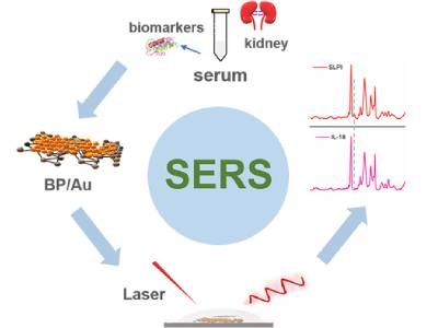
Photo: The new SERS spectroscopy method utilizes a gold and black phosphorous substrate and is used to detect two biomarkers associated with kidney injury in donor blood serum samples. Credit: Hui Chen, University of Shanghai for Science and Technology, and Mingxing Sui, Changhai Hospital of Shanghai